Copyright
©The Author(s) 2024.
World J Gastrointest Surg. Aug 27, 2024; 16(8): 2724-2734
Published online Aug 27, 2024. doi: 10.4240/wjgs.v16.i8.2724
Published online Aug 27, 2024. doi: 10.4240/wjgs.v16.i8.2724
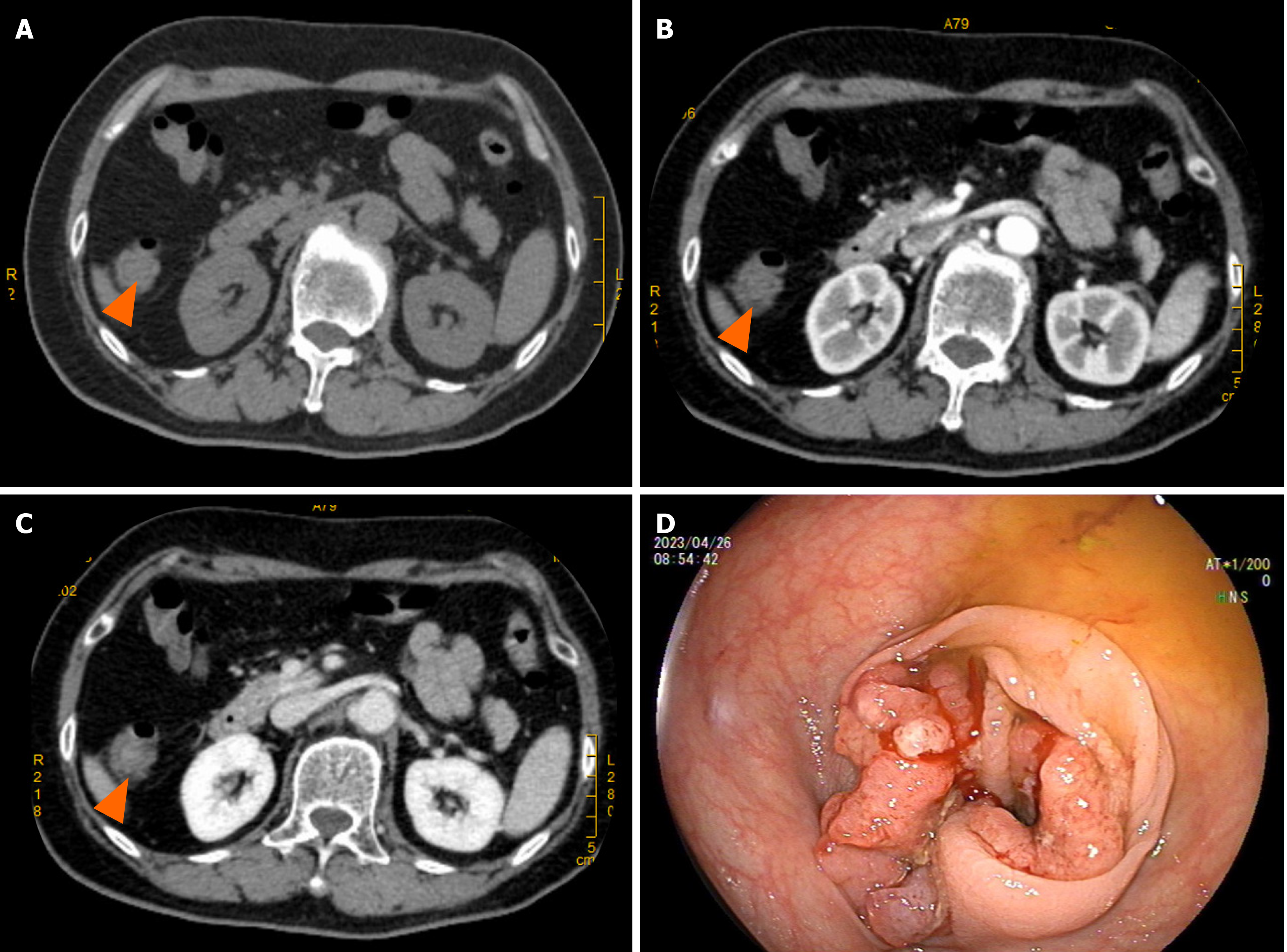
Figure 1 Colonoscopy and enhanced total abdominal computed tomography.
A-C: Computed tomography (CT) shows that the colon cancer near the hepatic flexure of the transverse colon invaded the serosal layer, with a maximum invasion depth of approximately 2 cm, with small mesenteric lymph node metastasis (orange triangles) to be ruled out; CT plain scan (A), CT enhanced scan (B), and CT venous phase of the mass (C), respectively; D: Colonoscopy shows an irregular mass with a diameter of > 2 cm located 70 cm from the anus.
- Citation: Fei S, Wu WD, Zhang HS, Liu SJ, Li D, Jin B. Primary coexisting adenocarcinoma of the colon and neuroendocrine tumor of the duodenum: A case report and review of the literature. World J Gastrointest Surg 2024; 16(8): 2724-2734
- URL: https://www.wjgnet.com/1948-9366/full/v16/i8/2724.htm
- DOI: https://dx.doi.org/10.4240/wjgs.v16.i8.2724


