Copyright
©The Author(s) 2024.
World J Gastrointest Surg. Aug 27, 2024; 16(8): 2602-2611
Published online Aug 27, 2024. doi: 10.4240/wjgs.v16.i8.2602
Published online Aug 27, 2024. doi: 10.4240/wjgs.v16.i8.2602
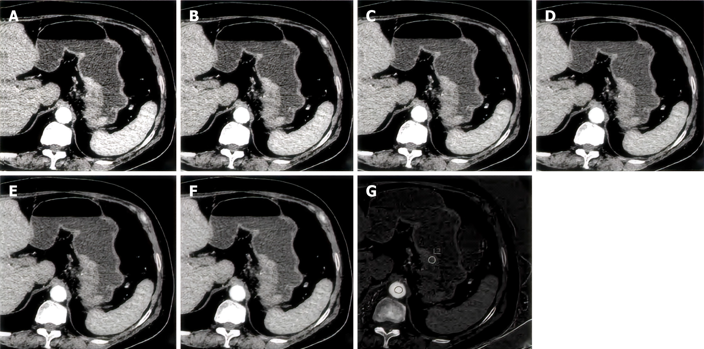
Figure 1 A case of PNI in a 76-year-old elderly woman with gastric cancer.
The tumor was located on the small curved side, and the region of interest was 110 mm2. A: Computed tomography (CT)60 keV in single-energy images of the arterial phase; B: CT70 keV in single-energy images of the arterial phase; C: CT80 keV in single-energy images of the arterial phase; D: CT90 keV in single-energy images of the arterial phase; E: CT100 keV in single-energy images of the arterial phase; F: CT110 keV in single-energy images of the arterial phase; G: Iodine base map.
- Citation: Lan YY, Han J, Liu YY, Lan L. Construction of a predictive model for gastric cancer neuroaggression and clinical validation analysis: A single-center retrospective study. World J Gastrointest Surg 2024; 16(8): 2602-2611
- URL: https://www.wjgnet.com/1948-9366/full/v16/i8/2602.htm
- DOI: https://dx.doi.org/10.4240/wjgs.v16.i8.2602


