Copyright
©The Author(s) 2024.
World J Gastrointest Surg. Jul 27, 2024; 16(7): 2351-2357
Published online Jul 27, 2024. doi: 10.4240/wjgs.v16.i7.2351
Published online Jul 27, 2024. doi: 10.4240/wjgs.v16.i7.2351
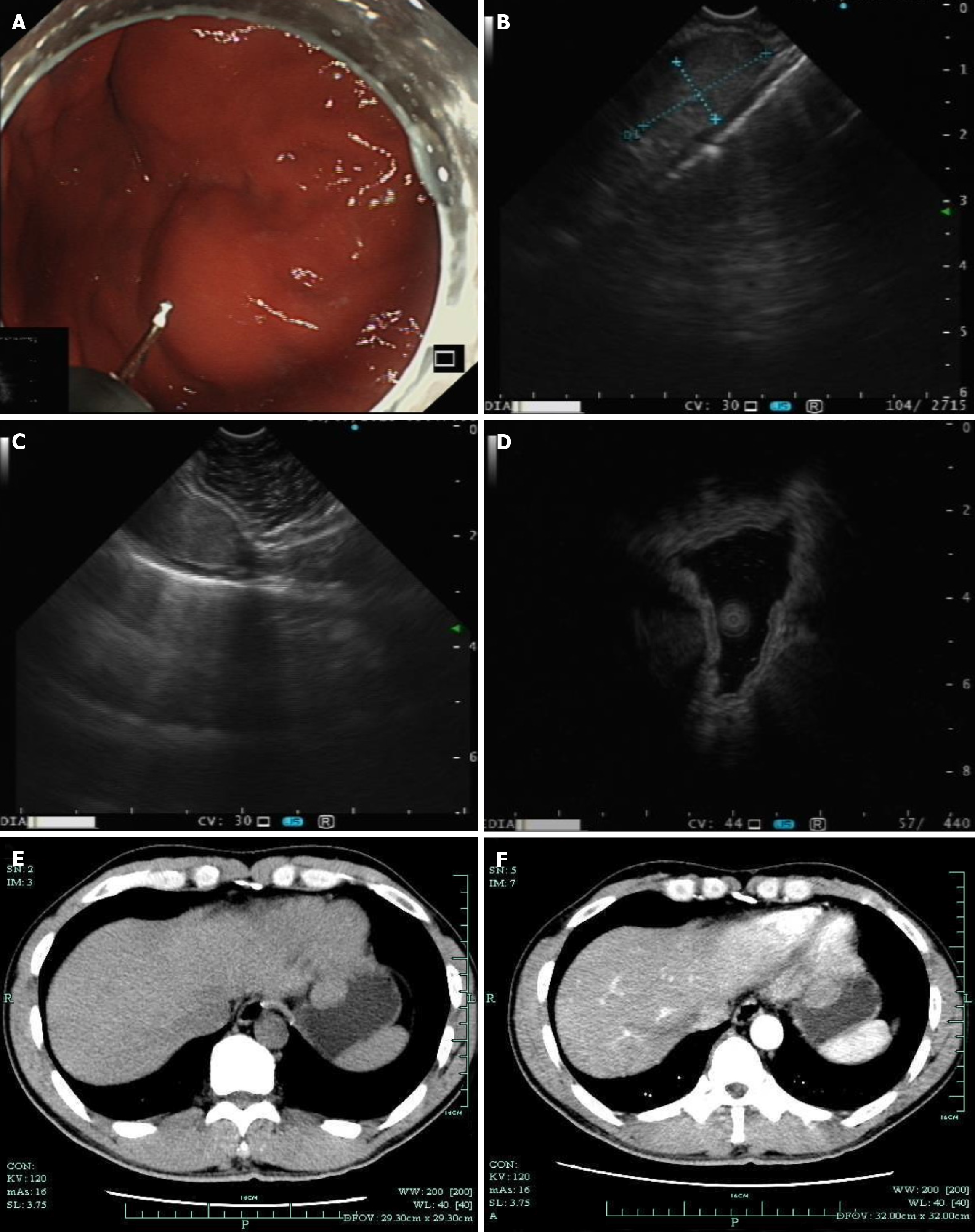
Figure 1 Imaging figures related to case 1.
A: During white-light gastroscopy, a submucosal mass of approximately 2 cm was visible at the gastric fundus, covered by the normal gastric mucosal epithelium; B and C: Under linear endoscopic ultrasound, the lesion appeared as a hypoechoic mass, suspected to originate from the muscularis propria layer. However, on the left side of the image, a complete 5-layer structure of the gastric wall was visible (C); D: Miniprobe sonography scanning of the lesion revealed a hypoechoic mass suspected to originate from the muscularis propria layer; E: During the computed tomography non-contrast phase, a protruding mass into the gastric lumen was visible; F: The computed tomography contrast-enhanced phase, the lesion showed uniform mild enhancement.
- Citation: Wang JJ, Zhang FM, Chen W, Zhu HT, Gui NL, Li AQ, Chen HT. Misdiagnosis of hemangioma of left triangular ligament of the liver as gastric submucosal stromal tumor: Two case reports. World J Gastrointest Surg 2024; 16(7): 2351-2357
- URL: https://www.wjgnet.com/1948-9366/full/v16/i7/2351.htm
- DOI: https://dx.doi.org/10.4240/wjgs.v16.i7.2351


