Copyright
©The Author(s) 2024.
World J Gastrointest Surg. Jul 27, 2024; 16(7): 2329-2336
Published online Jul 27, 2024. doi: 10.4240/wjgs.v16.i7.2329
Published online Jul 27, 2024. doi: 10.4240/wjgs.v16.i7.2329
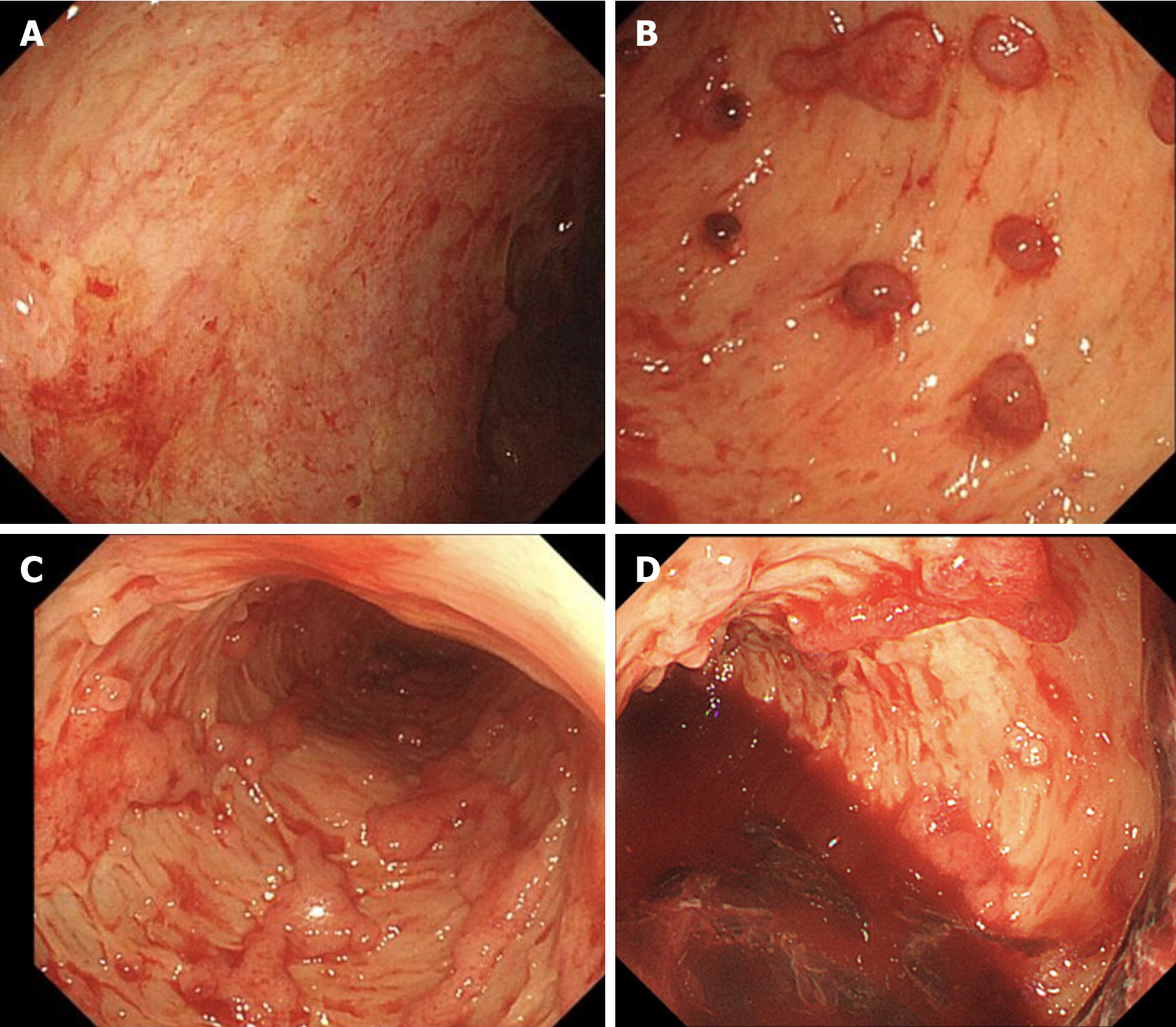
Figure 2 Sigmoidoscopy examination.
A: The lesions in the rectum, which were less than 5 cm, appeared to be relatively mild; B and C: Extensive mucosal exfoliation was observed below 30 cm in the sigmoid colon, along with scattered, small, dotted mucous membranes; D: A significant presence of blood and clots was found in the sigmoid colon.
- Citation: Hong N, Wang B, Zhou HC, Wu ZX, Fang HY, Song GQ, Yu Y. Multidisciplinary management of ulcerative colitis complicated by immune checkpoint inhibitor-associated colitis with life-threatening gastrointestinal hemorrhage: A case report. World J Gastrointest Surg 2024; 16(7): 2329-2336
- URL: https://www.wjgnet.com/1948-9366/full/v16/i7/2329.htm
- DOI: https://dx.doi.org/10.4240/wjgs.v16.i7.2329


