Copyright
©The Author(s) 2024.
World J Gastrointest Surg. Jul 27, 2024; 16(7): 2135-2144
Published online Jul 27, 2024. doi: 10.4240/wjgs.v16.i7.2135
Published online Jul 27, 2024. doi: 10.4240/wjgs.v16.i7.2135
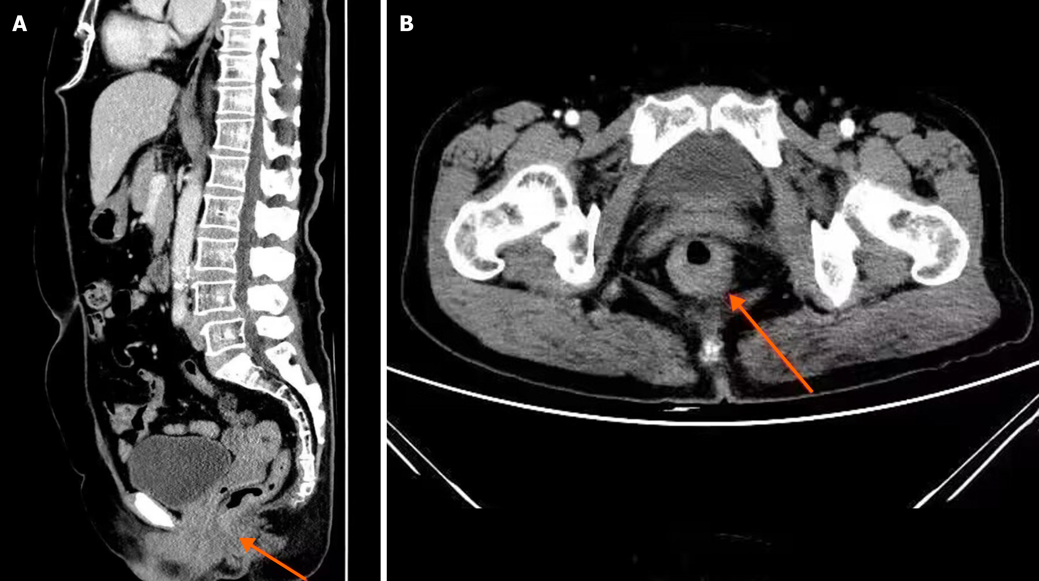
Figure 1 Imaging images.
A: Sagittal image; B: Cross-sectional image. The orange arrows show that the lower rectal wall of the patient was thickened, and obvious changes were observed after enhancement. Small lymph nodes were seen around the lesion. The sample images were from the digital medical record database.
- Citation: Bai LN, Zhang LX. Effectiveness of magnetic resonance imaging and spiral computed tomography in the staging and treatment prognosis of colorectal cancer. World J Gastrointest Surg 2024; 16(7): 2135-2144
- URL: https://www.wjgnet.com/1948-9366/full/v16/i7/2135.htm
- DOI: https://dx.doi.org/10.4240/wjgs.v16.i7.2135


