Copyright
©The Author(s) 2024.
World J Gastrointest Surg. Jul 27, 2024; 16(7): 1986-2002
Published online Jul 27, 2024. doi: 10.4240/wjgs.v16.i7.1986
Published online Jul 27, 2024. doi: 10.4240/wjgs.v16.i7.1986
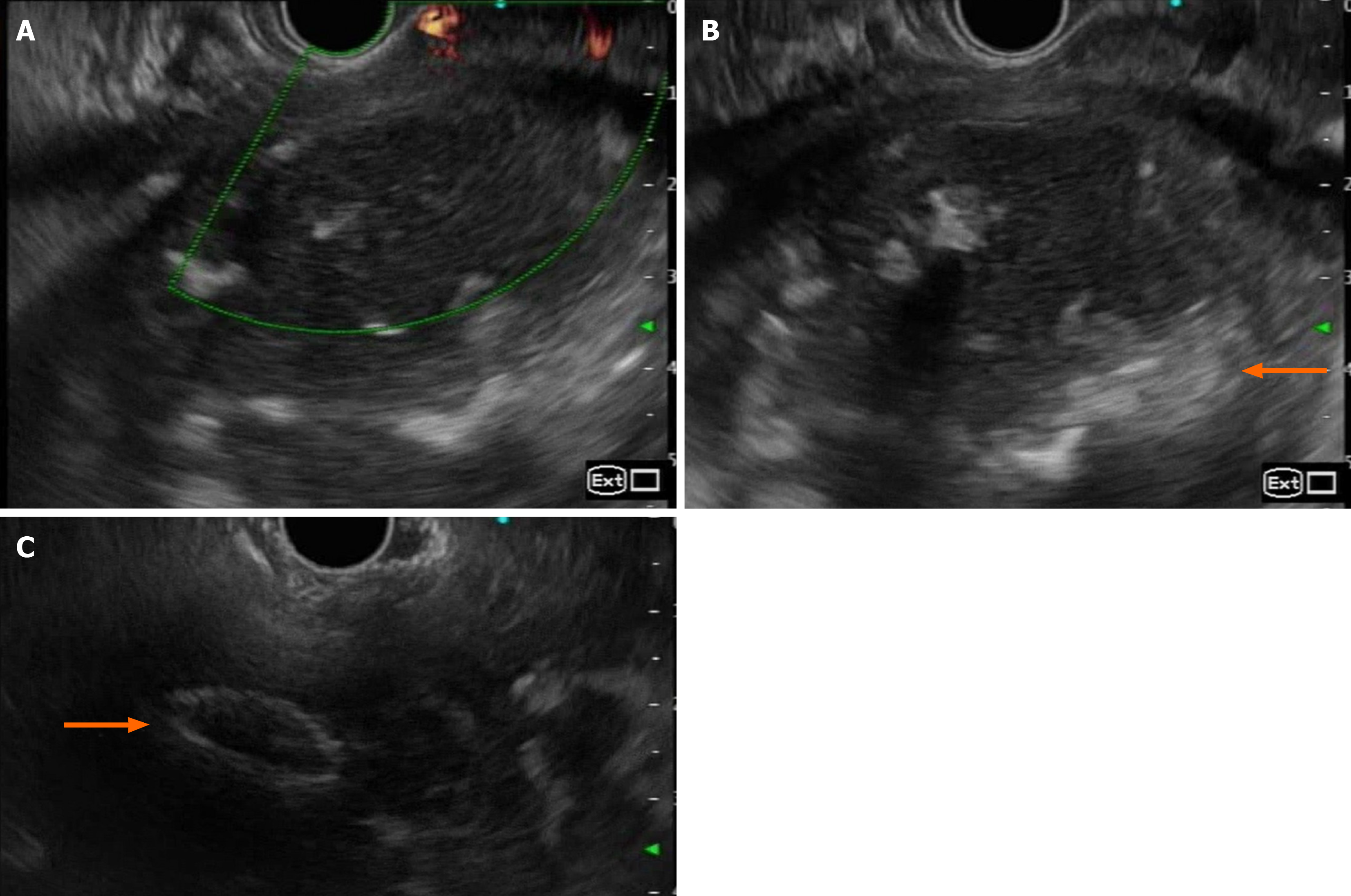
Figure 7 Endoscopic ultrasound images.
A: Absence of Doppler signal within walled-off necrosis cavity, ruling out presence of vessels or pseudoaneurysm; B: Solid debris (labelled with arrow) seen within walled-off necrosis cavity; C: Post deployment of lumen-apposing metal stent (labelled with arrow).
- Citation: Koo JG, Liau MYQ, Kryvoruchko IA, Habeeb TA, Chia C, Shelat VG. Pancreatic pseudocyst: The past, the present, and the future. World J Gastrointest Surg 2024; 16(7): 1986-2002
- URL: https://www.wjgnet.com/1948-9366/full/v16/i7/1986.htm
- DOI: https://dx.doi.org/10.4240/wjgs.v16.i7.1986


