Copyright
©The Author(s) 2024.
World J Gastrointest Surg. Jun 27, 2024; 16(6): 1918-1925
Published online Jun 27, 2024. doi: 10.4240/wjgs.v16.i6.1918
Published online Jun 27, 2024. doi: 10.4240/wjgs.v16.i6.1918
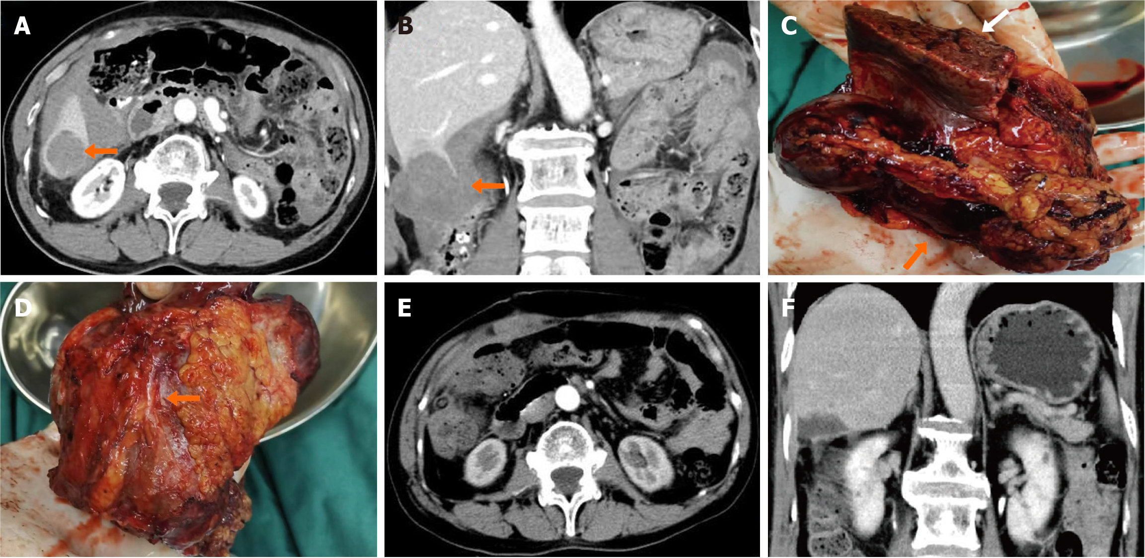
Figure 1 Computed tomography imaging before and after the first surgery.
A: Axial view of the tumor prior to surgery; B: Coronal view of the tumor prior to surgery; C: Gross specimen of surgically excised neoplastic lesion. The arrow indicates the margin of the liver and the tumor surrounding the liver; D: The arrow shows the adhesions between the tumor and the colon; E: Axial view after surgery; F: Coronal view after surgery.
- Citation: Zhang HL, Zhang M, Guo JQ, Wu FN, Zhu JD, Tu CY, Lv XL, Zhang K. Malignant myopericytoma originating from the colon: A case report. World J Gastrointest Surg 2024; 16(6): 1918-1925
- URL: https://www.wjgnet.com/1948-9366/full/v16/i6/1918.htm
- DOI: https://dx.doi.org/10.4240/wjgs.v16.i6.1918


