Copyright
©The Author(s) 2024.
World J Gastrointest Surg. Jun 27, 2024; 16(6): 1691-1699
Published online Jun 27, 2024. doi: 10.4240/wjgs.v16.i6.1691
Published online Jun 27, 2024. doi: 10.4240/wjgs.v16.i6.1691
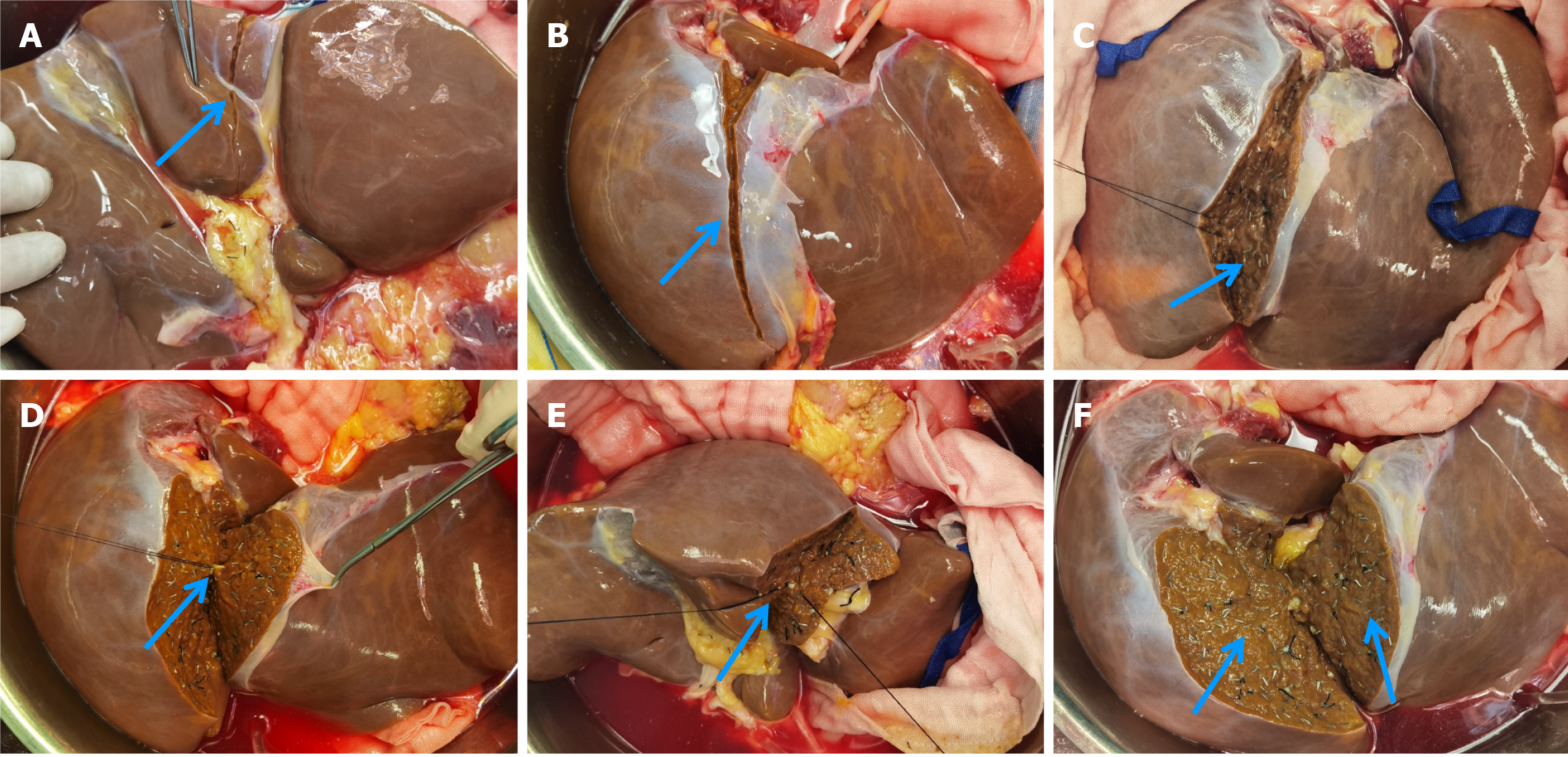
Figure 3 Procedure of liver parenchymal division.
A: Identification of the landmark line on the visceral surface of the liver for liver parenchymal division (0.5-1.0 cm to the right of the liver round ligament); B: Landmark line on the diaphragmatic surface of the liver for liver parenchymal division (on the right of the falciform ligament); C: Liver parenchymal division in the flat position, using titanium clips for small vessels (arrow); D: Adjusting the position of the liver during parenchymal division (suprahepatic inferior vena cava facing upwards), using silk sutures or ligatures for larger vessels (arrow); E: Continued liver parenchymal division with the liver flipped (suprahepatic inferior vena cava facing downward); F: Display of the two smooth liver segment surfaces after completion of the splitting process (arrow).
- Citation: Zhao D, Xie QH, Fang TS, Zhang KJ, Tang JX, Yan X, Jin X, Xie LJ, Xie WG. How to apply ex-vivo split liver transplantation safely and feasibly: A three-step approach. World J Gastrointest Surg 2024; 16(6): 1691-1699
- URL: https://www.wjgnet.com/1948-9366/full/v16/i6/1691.htm
- DOI: https://dx.doi.org/10.4240/wjgs.v16.i6.1691


