Copyright
©The Author(s) 2024.
World J Gastrointest Surg. Jun 27, 2024; 16(6): 1691-1699
Published online Jun 27, 2024. doi: 10.4240/wjgs.v16.i6.1691
Published online Jun 27, 2024. doi: 10.4240/wjgs.v16.i6.1691
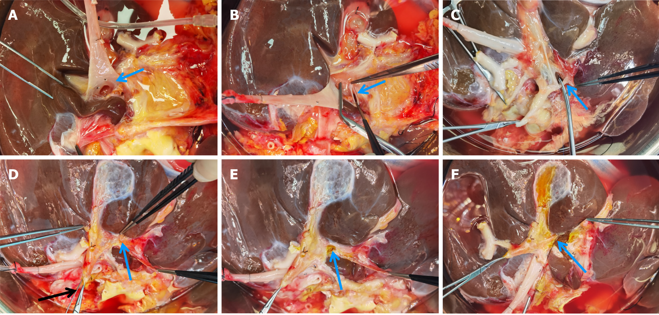
Figure 1 Procedure of splitting the first porta hepatis.
A: Separation of the left and right branches of the portal vein (arrow indicating the left portal vein); B: Division of the left portal vein (arrow) followed by suturing of the proximal end; C: Identification and division of the left hepatic artery (arrow); D: Identification of the division site of the left hepatic duct (blue arrow) under biliary probe guidance (black arrow); E: Incision of the left hepatic duct anterior wall (arrow) and reconfirmation of the left hepatic duct, right hepatic duct, and suspected bile duct openings using the probe; F: Division of the left hepatic duct, confirming the landmark for the division of liver parenchyma in the first porta hepatis.
- Citation: Zhao D, Xie QH, Fang TS, Zhang KJ, Tang JX, Yan X, Jin X, Xie LJ, Xie WG. How to apply ex-vivo split liver transplantation safely and feasibly: A three-step approach. World J Gastrointest Surg 2024; 16(6): 1691-1699
- URL: https://www.wjgnet.com/1948-9366/full/v16/i6/1691.htm
- DOI: https://dx.doi.org/10.4240/wjgs.v16.i6.1691


