Copyright
©The Author(s) 2024.
World J Gastrointest Surg. May 27, 2024; 16(5): 1449-1460
Published online May 27, 2024. doi: 10.4240/wjgs.v16.i5.1449
Published online May 27, 2024. doi: 10.4240/wjgs.v16.i5.1449
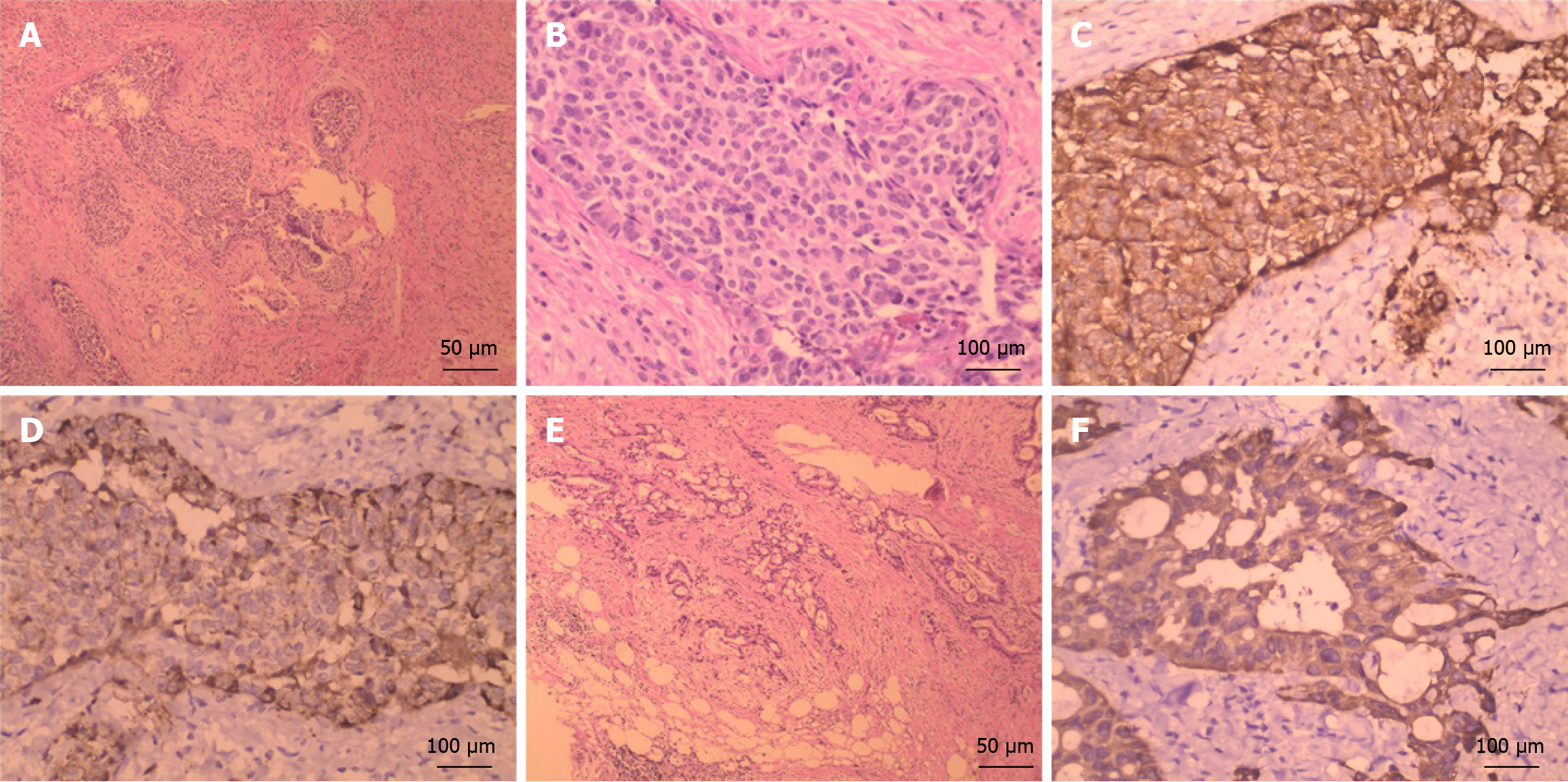
Figure 2 Histopathologic features in neuroendocrine carcinoma of the common hepatic duct coexisting with distal cholangiocarcinoma.
A: The neuroendocrine carcinoma (NEC) showed a nested organoid growth pattern [hematoxylin and eosin (HE), × 100]; B: The NEC cells were round or oval, hyperchromatic nuclei and scant cytoplasm (HE, × 400); C: Immunohistochemically, the NEC cells were positive for synaptophysin(HE, × 400); D: Immunohistochemically, the NEC cells were positive for chromogranin A (HE, × 400); E: The distal cholangiocarcinoma (dCCA) cells arranged in irregular tubular and papillary structures (HE, × 100); F: Immunohistochemically, the dCCA cells were positive for cytokeratin 19 (HE, × 400).
- Citation: Chen F, Li WW, Mo JF, Chen MJ, Wang SH, Yang SY, Song ZW. Neuroendocrine carcinoma of the common hepatic duct coexisting with distal cholangiocarcinoma: A case report and review of literature. World J Gastrointest Surg 2024; 16(5): 1449-1460
- URL: https://www.wjgnet.com/1948-9366/full/v16/i5/1449.htm
- DOI: https://dx.doi.org/10.4240/wjgs.v16.i5.1449


