Copyright
©The Author(s) 2024.
World J Gastrointest Surg. May 27, 2024; 16(5): 1430-1435
Published online May 27, 2024. doi: 10.4240/wjgs.v16.i5.1430
Published online May 27, 2024. doi: 10.4240/wjgs.v16.i5.1430
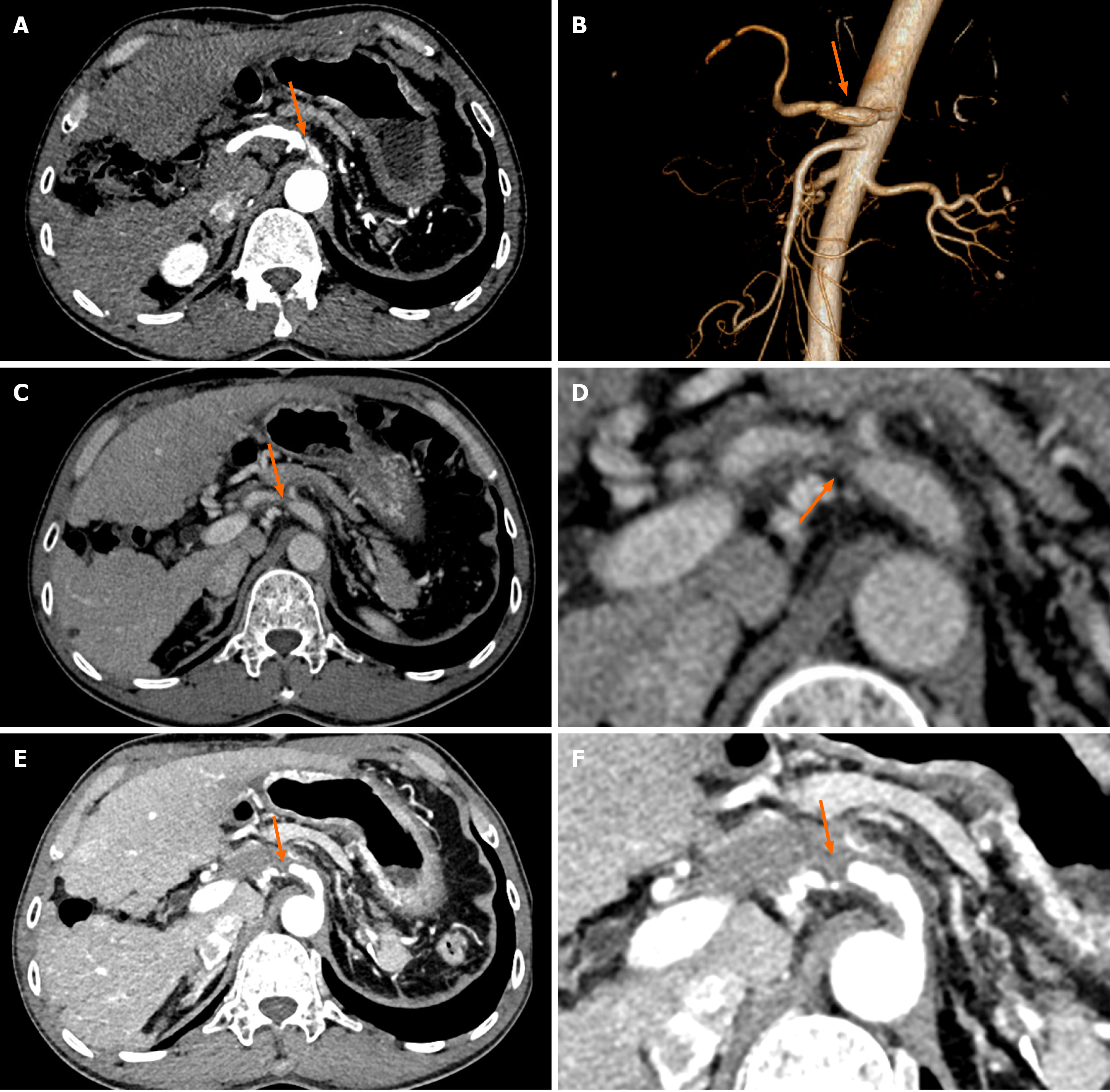
Figure 2 Contrast-enhanced computed tomography images of the patient.
A and B: Artery dissection is shown in the first computed tomography examination (orange arrow); C and D: Narrowing of the common hepatic artery at the 3-month follow-up (orange arrow); E and F: Occlusion of the common hepatic artery with collateralization at the 6-month follow-up (orange arrow).
- Citation: Pu Y, Luo Y. Multi-modal imaging for the diagnosis of spontaneous visceral artery dissection: A case report. World J Gastrointest Surg 2024; 16(5): 1430-1435
- URL: https://www.wjgnet.com/1948-9366/full/v16/i5/1430.htm
- DOI: https://dx.doi.org/10.4240/wjgs.v16.i5.1430


