Copyright
©The Author(s) 2024.
World J Gastrointest Surg. May 27, 2024; 16(5): 1385-1394
Published online May 27, 2024. doi: 10.4240/wjgs.v16.i5.1385
Published online May 27, 2024. doi: 10.4240/wjgs.v16.i5.1385
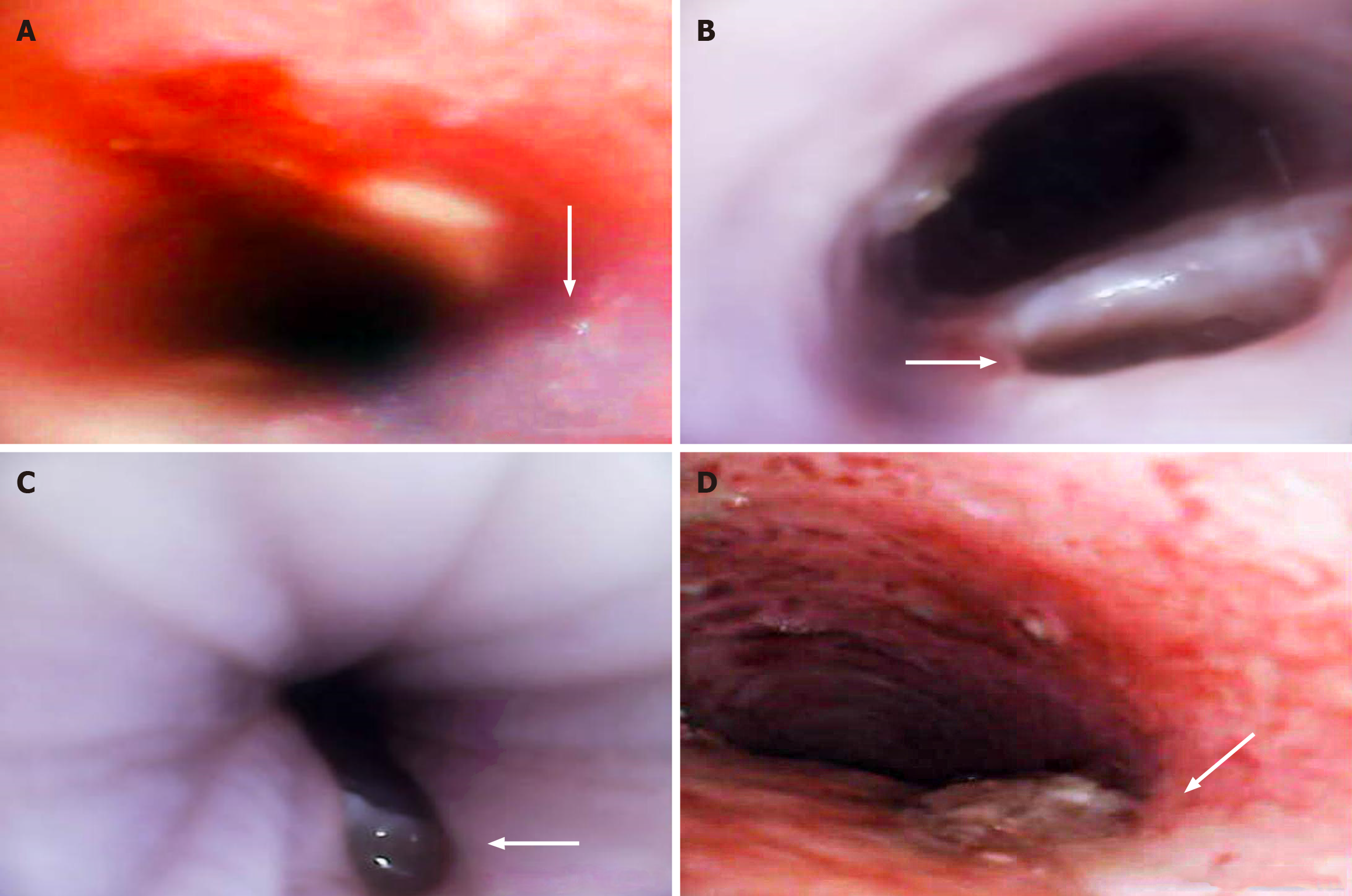
Figure 3 Endoscopic findings after modeling with the surgical modeling technique and modified magnetic compression technique.
A: In the surgical modeling (SM) group, methylene blue was immediate injected into the esophagus, leading to overflow into the trachea (indicated by the arrow); B: 5 d after successful modeling with modified magnetic compression (MMC) technique, the fistula on the tracheal side was observed (indicated by the arrow); C: 5 d after successful modeling with MMC technique, the fistula on the esophageal side was observed (indicated by the arrow); D: 5 d after successful modeling with SM, the fistula and surrounding tracheal mucosa were observed (indicated by the arrow).
- Citation: Meng H, Nan FY, Kou N, Hong QY, Lv MS, Li JB, Zhang BJ, Zou H, Li L, Wang HW. Establishment of acquired tracheoesophageal fistula using a modified magnetic compression technique in rabbits and its postmodeling evaluation. World J Gastrointest Surg 2024; 16(5): 1385-1394
- URL: https://www.wjgnet.com/1948-9366/full/v16/i5/1385.htm
- DOI: https://dx.doi.org/10.4240/wjgs.v16.i5.1385


