Copyright
©The Author(s) 2024.
World J Gastrointest Surg. Apr 27, 2024; 16(4): 1195-1202
Published online Apr 27, 2024. doi: 10.4240/wjgs.v16.i4.1195
Published online Apr 27, 2024. doi: 10.4240/wjgs.v16.i4.1195
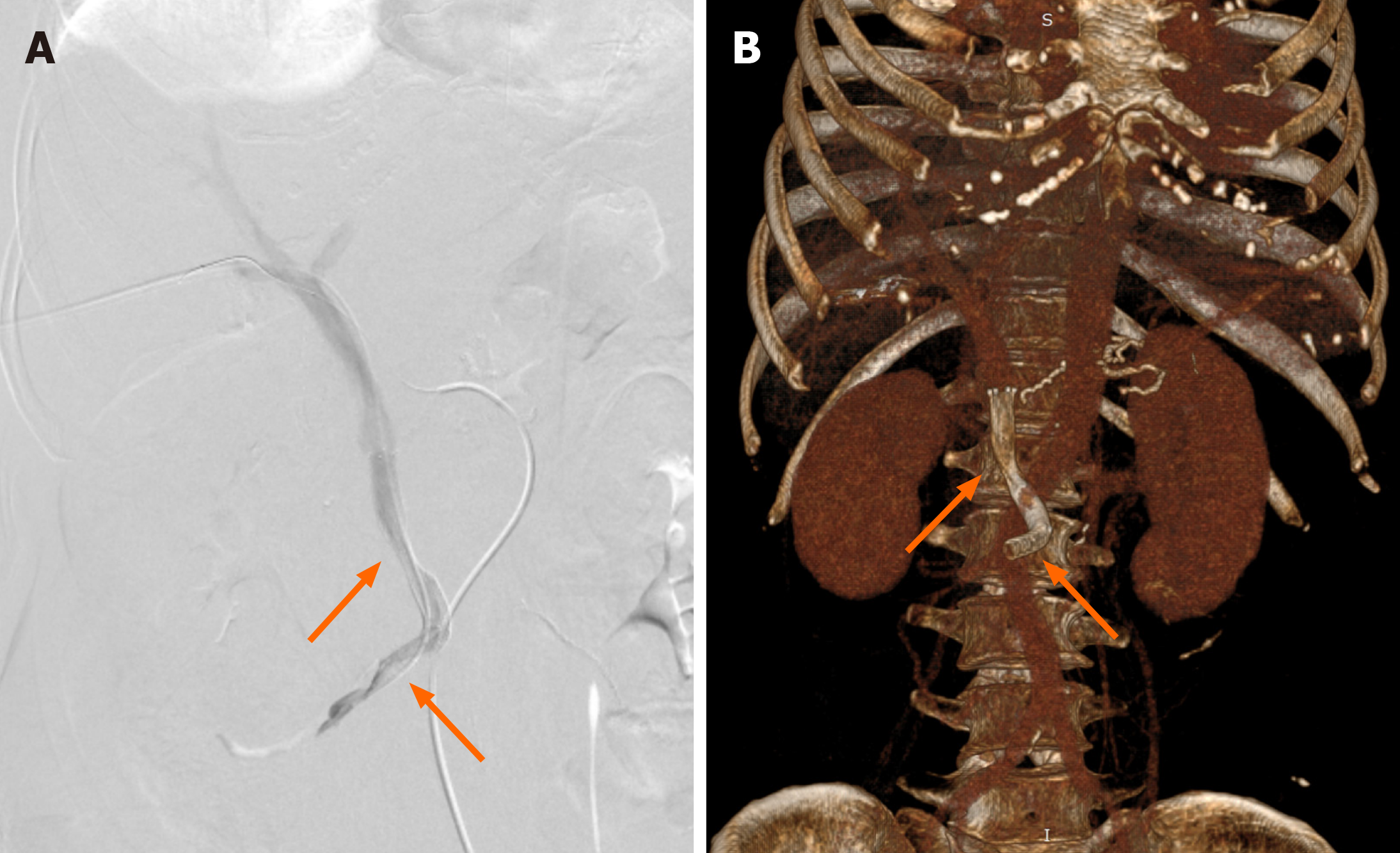
Figure 3 Percutaneous transhepatic direct portography showing improved stenosis after placement of two stents (arrows).
A: Digital subtraction angiography of portal vein; B: Volume rendering images showing stents in superior mesenteric vein/portal vein. Stent parameters: Upper arrow: 8 mm × 50 mm, coated; lower arrow: 8 mm × 60 mm, uncoated.
- Citation: Lin C, Wang ZY, Dong LB, Wang ZW, Li ZH, Wang WB. Percutaneous transhepatic stenting for acute superior mesenteric vein stenosis after pancreaticoduodenectomy with portal vein reconstruction: A case report. World J Gastrointest Surg 2024; 16(4): 1195-1202
- URL: https://www.wjgnet.com/1948-9366/full/v16/i4/1195.htm
- DOI: https://dx.doi.org/10.4240/wjgs.v16.i4.1195


