Copyright
©The Author(s) 2024.
World J Gastrointest Surg. Apr 27, 2024; 16(4): 1189-1194
Published online Apr 27, 2024. doi: 10.4240/wjgs.v16.i4.1189
Published online Apr 27, 2024. doi: 10.4240/wjgs.v16.i4.1189
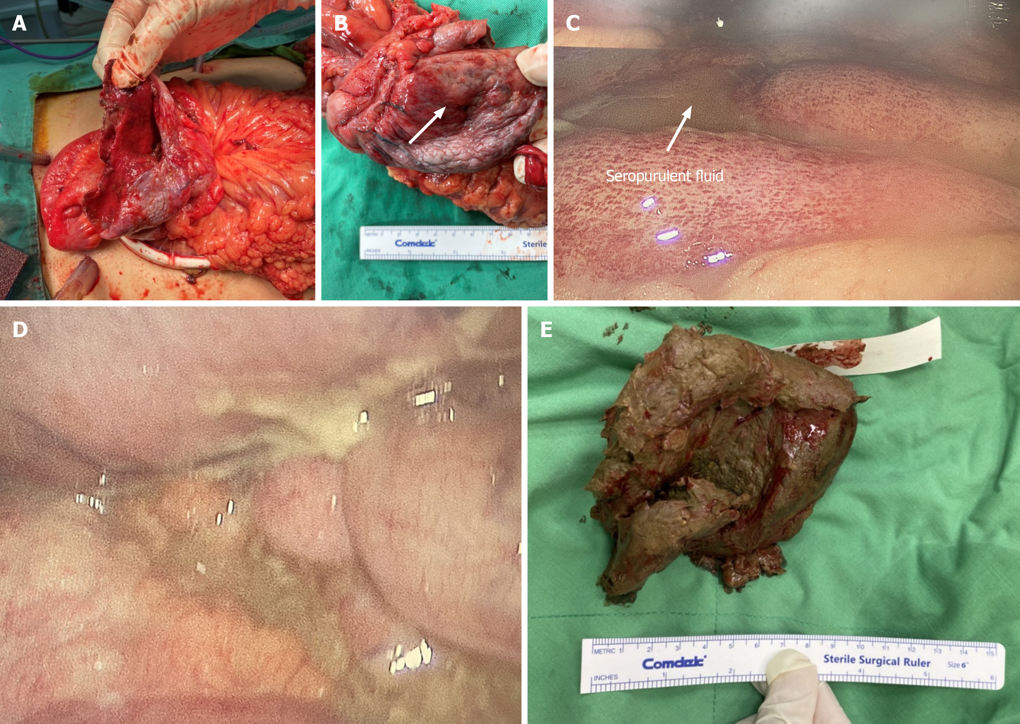
Figure 3 Intraoperative photographs of gross findings.
A: A rounded perforation (7 cm × 5 cm in diameter) of the cecum was intraoperatively noted; B: The cecum was notably thin without any obvious tumorous lesions on the mucosa (as marked); C: Seropurulent fluid was freely present in the peritoneal cavity; D: Fecal contamination was observed in the peritoneal cavity; E: The bezoar extracted from the dilated cecum.
- Citation: Yu HC, Pu TW, Kang JC, Chen CY, Hu JM, Su RY. Stercoral perforation of the cecum: A case report. World J Gastrointest Surg 2024; 16(4): 1189-1194
- URL: https://www.wjgnet.com/1948-9366/full/v16/i4/1189.htm
- DOI: https://dx.doi.org/10.4240/wjgs.v16.i4.1189


