Copyright
©The Author(s) 2024.
World J Gastrointest Surg. Apr 27, 2024; 16(4): 1066-1077
Published online Apr 27, 2024. doi: 10.4240/wjgs.v16.i4.1066
Published online Apr 27, 2024. doi: 10.4240/wjgs.v16.i4.1066
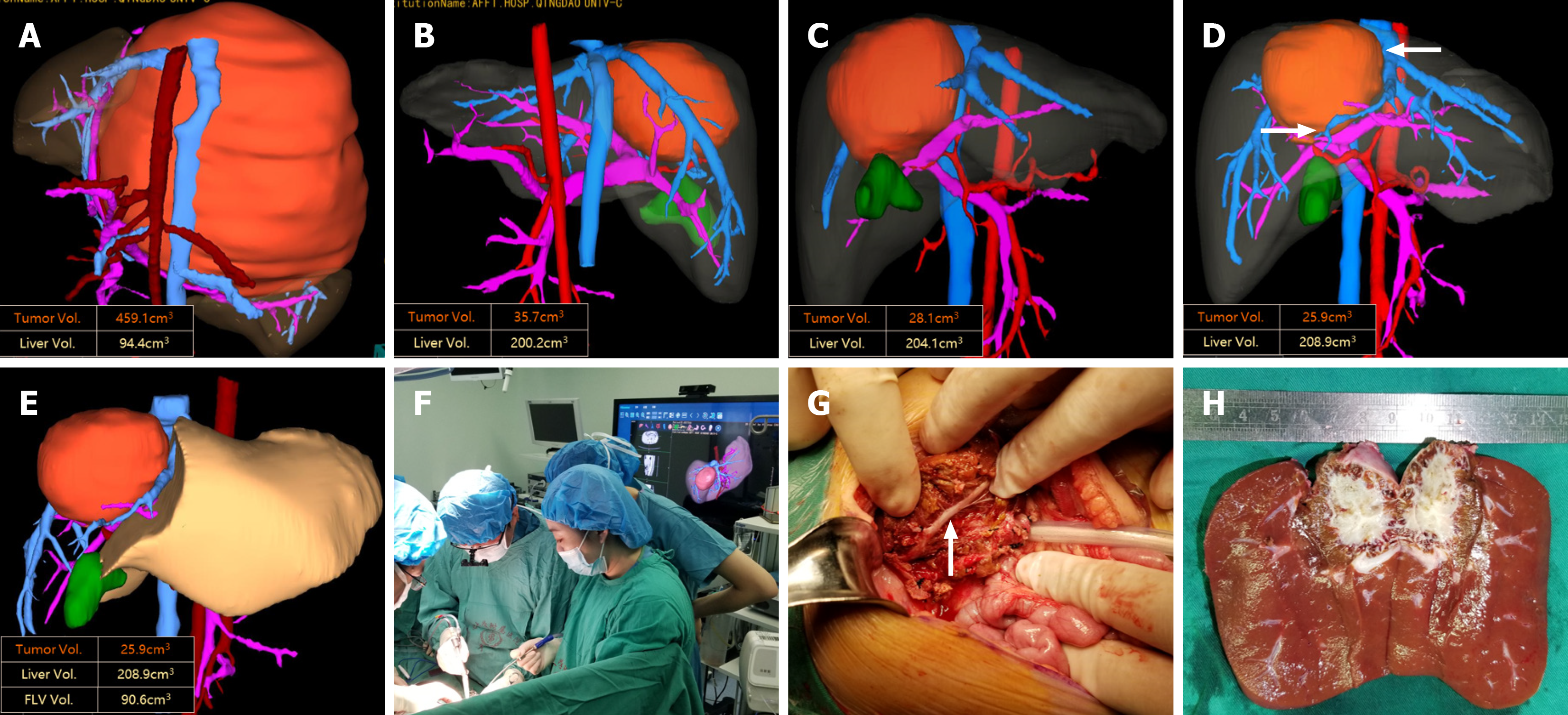
Figure 2 A case of giant hepatoblastoma in the middle lobe of the liver.
A: The tumor was initially discovered to be giant, causing distortion and displacement of all the intrahepatic blood vessels. A needle biopsy confirmed the presence of a hepatoblastoma, and neoadjuvant chemotherapy was recommended as the only viable option, in addition to liver transplantation; B: After 4 cycles of chemotherapy, the tumor volume was significantly reduced, but all intrahepatic blood vessels were compressed, making the tumor unresectable; C: Upon re-evaluation after 5 cycles, there was a slight reduction in tumor volume, but the tumor still remained in close proximity to major blood vessels of the liver, preventing surgical resection; D: After 6 cycles, there was no significant change in tumor volume. The tumor was touching the inferior vena cava and portal vein bifurcation, and surgical resection was considered; E: Preoperative simulation of right hemihepatectomy was conducted; F: Intraoperative three-dimensional images were used to assist real-time guidance during the right hemihepatectomy procedure; G and H: The operation proceeded successfully as the preoperative planning, resulting in complete tumor resection while preserving the middle hepatic vein.
- Citation: Xiu WL, Liu J, Zhang JL, Wang JM, Wang XF, Wang FF, Mi J, Hao XW, Xia N, Dong Q. Computer-assisted three-dimensional individualized extreme liver resection for hepatoblastoma in proximity to the major liver vasculature. World J Gastrointest Surg 2024; 16(4): 1066-1077
- URL: https://www.wjgnet.com/1948-9366/full/v16/i4/1066.htm
- DOI: https://dx.doi.org/10.4240/wjgs.v16.i4.1066


