Copyright
©The Author(s) 2024.
World J Gastrointest Surg. Mar 27, 2024; 16(3): 944-954
Published online Mar 27, 2024. doi: 10.4240/wjgs.v16.i3.944
Published online Mar 27, 2024. doi: 10.4240/wjgs.v16.i3.944
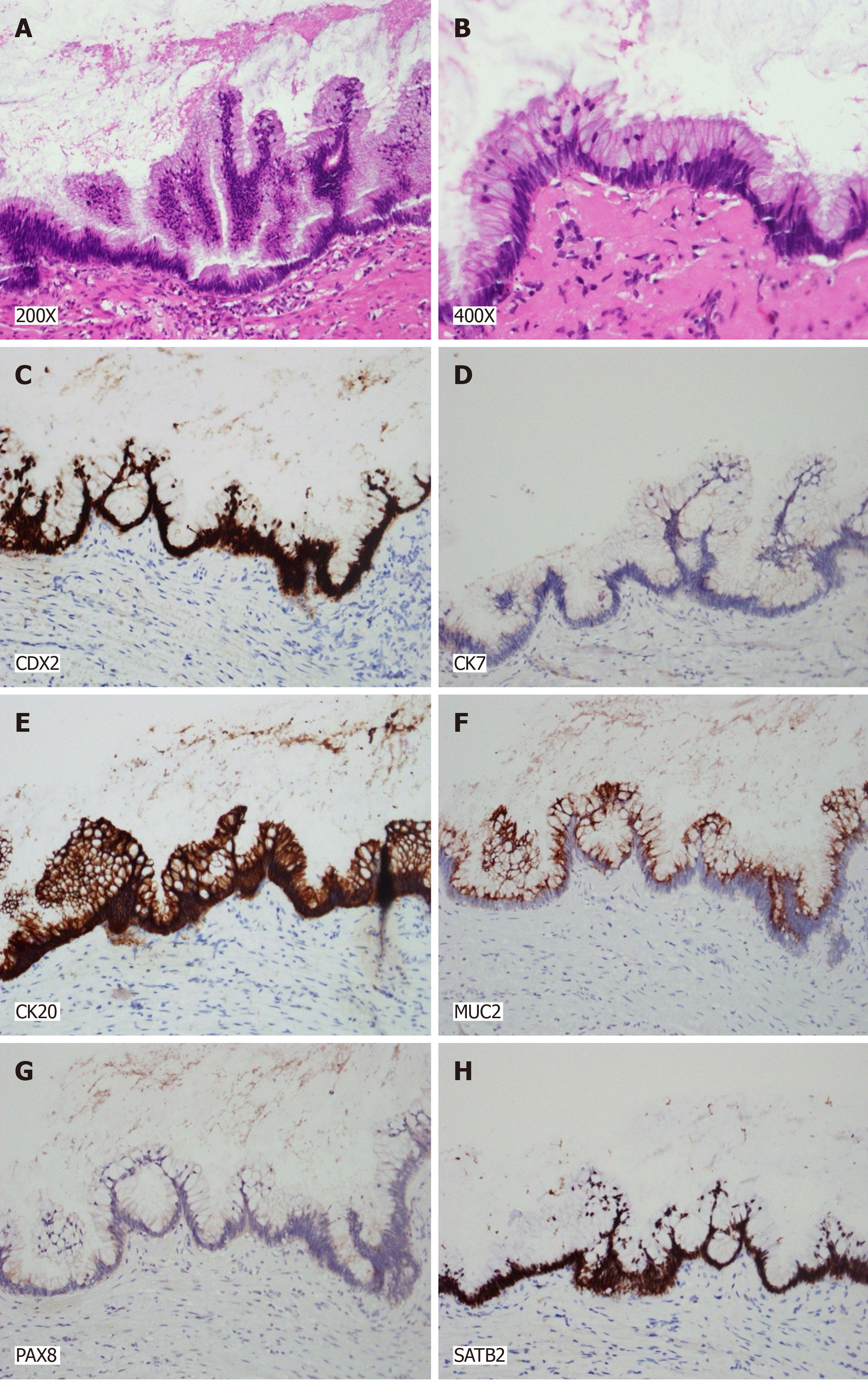
Figure 6 Histological examination of the lesion revealing characteristic patterns.
A: A 200 × magnification image showing slender villi lined by tall mucinous epithelial cells with low-grade dysplasia; B: A 400 × magnification image demonstrating tall mucinous epithelial cells with low-grade dysplasia, set within a fibrous stromal framework; C: Immunohistochemical staining displaying positive CDX2 expression in the villous cells; D: Immunohistochemical staining displaying positive CDX20 expression in the villous cells; E: Immunohistochemical staining displaying positive SATB2 expression in the villous cells; F: Immunohistochemical staining displaying positive MUC2 expression in the villous cells; G: Immunohistochemical staining showing the villous cells negative for CK7; H: Immunohistochemical staining showing the villous cells negative for PAX8.Collective histopathological and immunohistochemical findings indicate the lesion to be a low-grade appendiceal mucinous neoplasm. The evaluation of surgical margins shows no neoplastic presence, indicating a clear disease-free status.
- Citation: Chang HC, Kang JC, Pu TW, Su RY, Chen CY, Hu JM. Mucinous neoplasm of the appendix: A case report and review of literature. World J Gastrointest Surg 2024; 16(3): 944-954
- URL: https://www.wjgnet.com/1948-9366/full/v16/i3/944.htm
- DOI: https://dx.doi.org/10.4240/wjgs.v16.i3.944


