Copyright
©The Author(s) 2024.
World J Gastrointest Surg. Mar 27, 2024; 16(3): 944-954
Published online Mar 27, 2024. doi: 10.4240/wjgs.v16.i3.944
Published online Mar 27, 2024. doi: 10.4240/wjgs.v16.i3.944
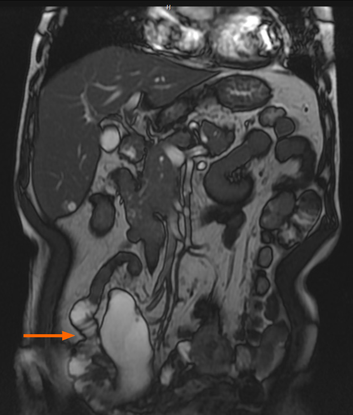
Figure 5 Preoperative coronal 2D Fast Imaging Employing Steady-state Acquisition magnetic resonance imaging of the abdomen revealed a cystic structure measuring approximately 9 cm in the right mesentery.
The imaging was conducted with a 1.5 Tesla superconducting magnet and a phased-array body coil. The patient was positioned supine, with the imaging parameters set to a field of view of 40 cm, a slice thickness of 6 mm, and an interslice gap of 1.5 mm. This lesion was subsequently identified as an appendiceal mucinous neoplasm.
- Citation: Chang HC, Kang JC, Pu TW, Su RY, Chen CY, Hu JM. Mucinous neoplasm of the appendix: A case report and review of literature. World J Gastrointest Surg 2024; 16(3): 944-954
- URL: https://www.wjgnet.com/1948-9366/full/v16/i3/944.htm
- DOI: https://dx.doi.org/10.4240/wjgs.v16.i3.944


