Copyright
©The Author(s) 2024.
World J Gastrointest Surg. Mar 27, 2024; 16(3): 944-954
Published online Mar 27, 2024. doi: 10.4240/wjgs.v16.i3.944
Published online Mar 27, 2024. doi: 10.4240/wjgs.v16.i3.944
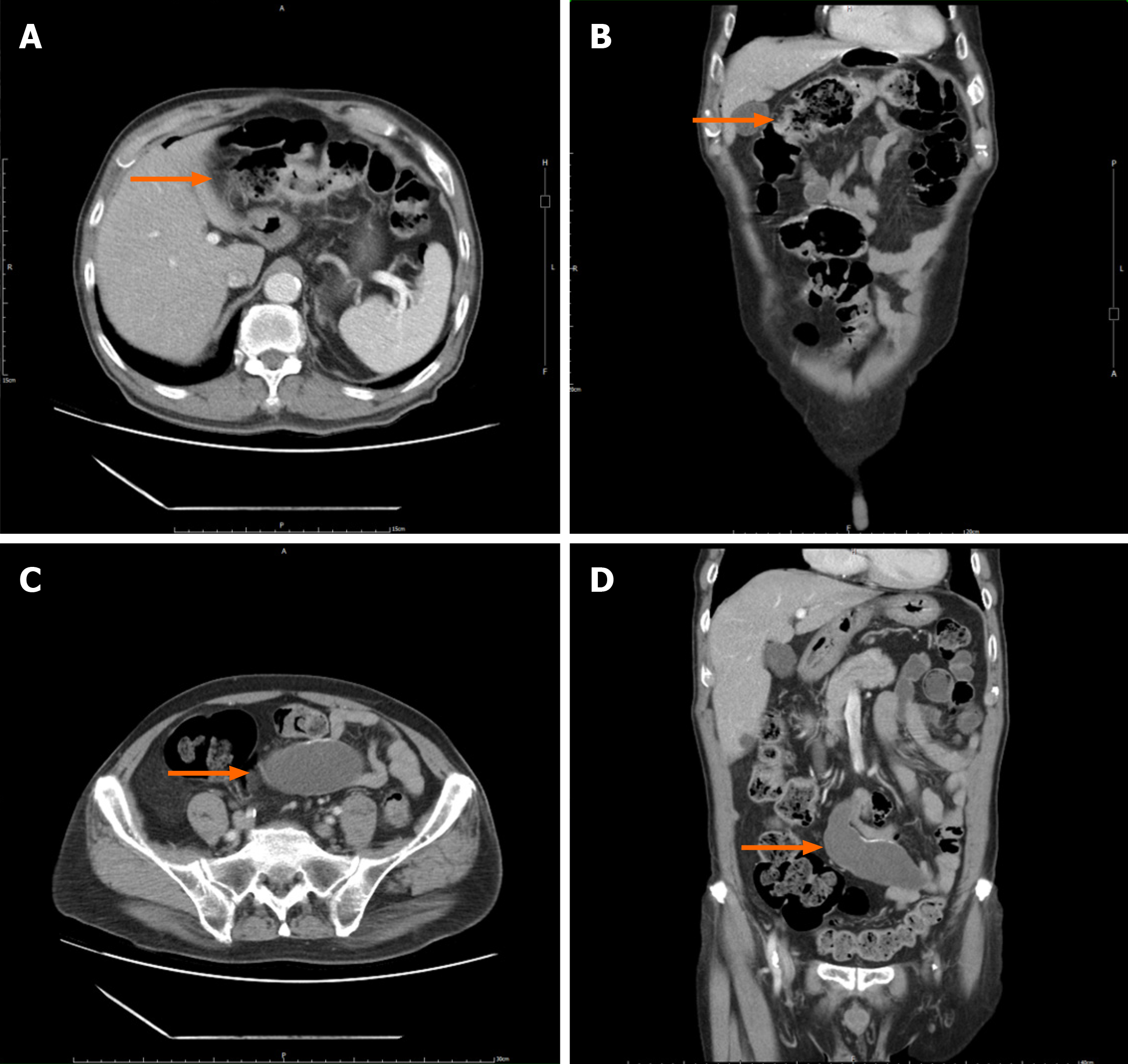
Figure 4 Preoperative abdominal computed tomography image.
A and B: The transverse colon wall and regional lymphadenopathy, correlating to a T3N1Mx stage, according to the 8th edition of the American Joint Committee on Cancer cancer staging guidelines (orange arrow); C and D: A cystic formation approximately 9 cm in size was initially detected and interpreted as a mesenteric cyst; subsequent scrutiny ascertained it as an appendiceal mucinous neoplasm (orange arrow).
- Citation: Chang HC, Kang JC, Pu TW, Su RY, Chen CY, Hu JM. Mucinous neoplasm of the appendix: A case report and review of literature. World J Gastrointest Surg 2024; 16(3): 944-954
- URL: https://www.wjgnet.com/1948-9366/full/v16/i3/944.htm
- DOI: https://dx.doi.org/10.4240/wjgs.v16.i3.944


