Copyright
©The Author(s) 2024.
World J Gastrointest Surg. Mar 27, 2024; 16(3): 944-954
Published online Mar 27, 2024. doi: 10.4240/wjgs.v16.i3.944
Published online Mar 27, 2024. doi: 10.4240/wjgs.v16.i3.944
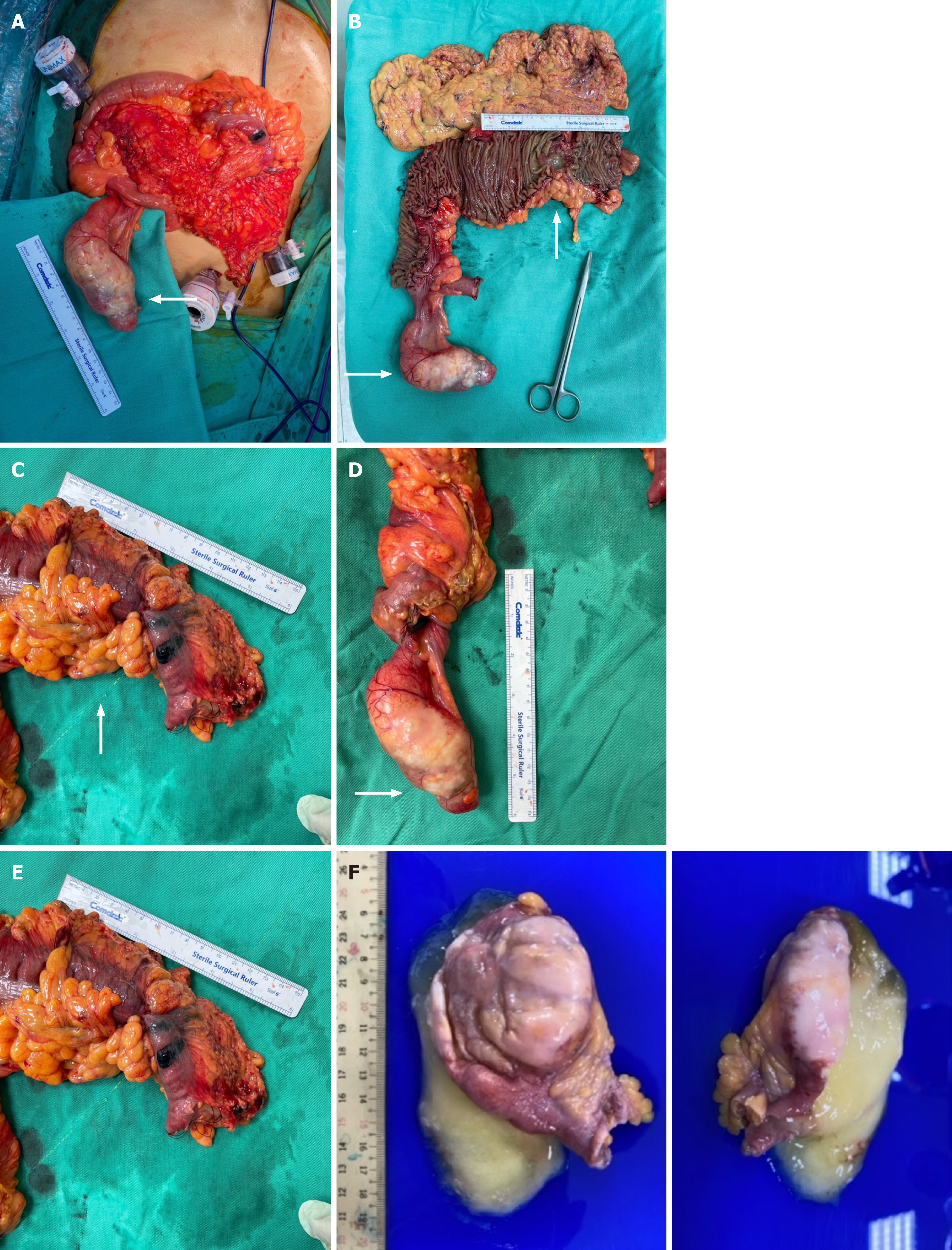
Figure 3 Multistage intraoperative and specimen images of colonic adenocarcinoma and appendiceal mucinous neoplasm.
A: Intraoperative photograph depicting the surgical field (white arrow); B: Comprehensive visualization of the adenocarcinoma and appendiceal mucinous neoplasm following an extended right hemicolectomy (white arrows); C: Image of the 6-cm neoplastic lesion located in the transverse colon (white arrow); D: Image of the 9-cm appendiceal mucinous neoplasm, featuring an intact capsular structure; E: Depiction of a large mucinous cavity within the appendix, which simulates a pseudo-diverticulum formation; F: The specimen submitted consists of one tissue fragment, measuring 10.6 cm × 4.0 cm × 3.3 cm in size, fixed in formalin. Grossly, it is opened and filled with mucoid materials.
- Citation: Chang HC, Kang JC, Pu TW, Su RY, Chen CY, Hu JM. Mucinous neoplasm of the appendix: A case report and review of literature. World J Gastrointest Surg 2024; 16(3): 944-954
- URL: https://www.wjgnet.com/1948-9366/full/v16/i3/944.htm
- DOI: https://dx.doi.org/10.4240/wjgs.v16.i3.944


