Copyright
©The Author(s) 2024.
World J Gastrointest Surg. Mar 27, 2024; 16(3): 944-954
Published online Mar 27, 2024. doi: 10.4240/wjgs.v16.i3.944
Published online Mar 27, 2024. doi: 10.4240/wjgs.v16.i3.944
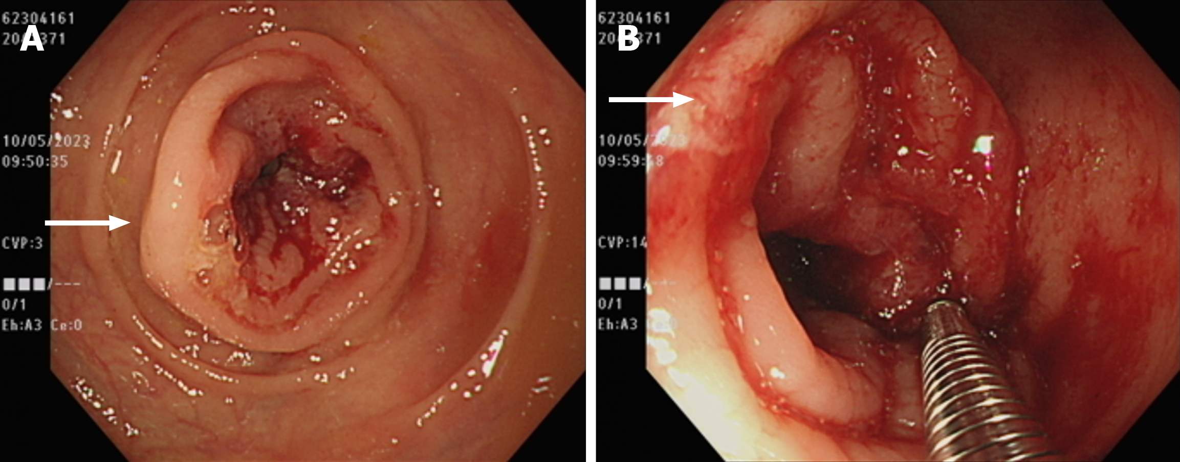
Figure 2 Diagnostic imaging and interventions with the colonoscope.
A: Colonoscopic examination revealed a 3-cm ulcerative lesion within the transverse colon, characterized by irregular luminal stenosis (white arrow); B: An attempt was made to obtain a biopsy through the colonoscope for pathological examination (white arrow).
- Citation: Chang HC, Kang JC, Pu TW, Su RY, Chen CY, Hu JM. Mucinous neoplasm of the appendix: A case report and review of literature. World J Gastrointest Surg 2024; 16(3): 944-954
- URL: https://www.wjgnet.com/1948-9366/full/v16/i3/944.htm
- DOI: https://dx.doi.org/10.4240/wjgs.v16.i3.944


