Copyright
©The Author(s) 2024.
World J Gastrointest Surg. Mar 27, 2024; 16(3): 932-943
Published online Mar 27, 2024. doi: 10.4240/wjgs.v16.i3.932
Published online Mar 27, 2024. doi: 10.4240/wjgs.v16.i3.932
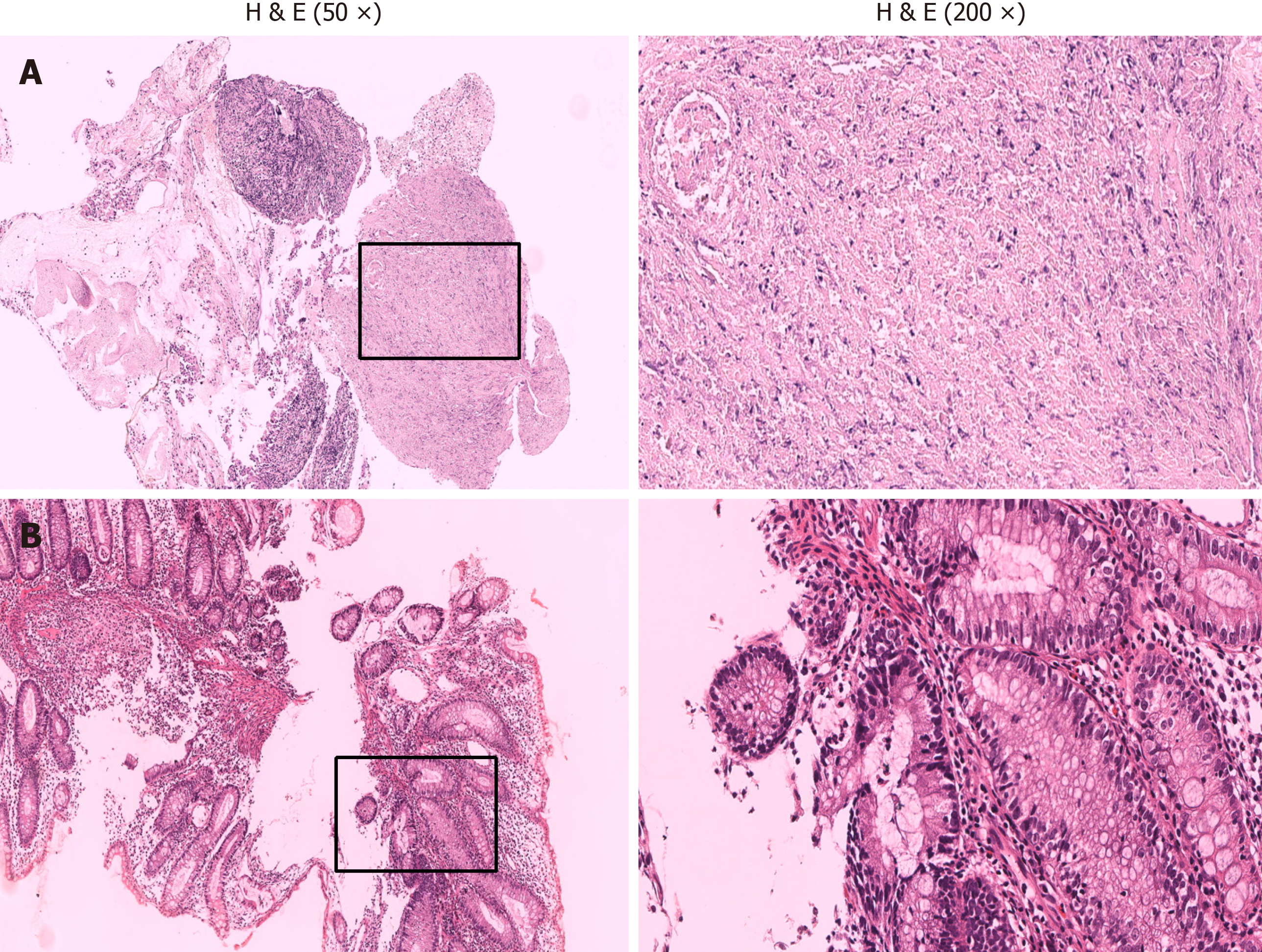
Figure 4 Pathology of intestinal mucosal tissue.
A: Colon (a small amount of mucosal tissue and inflammatory exudative necrosis); B: Distal ileum (ulcer formation, large numbers of inflammatory exudates and granuloma formation, and large numbers of lymphocytic plasma cell infiltration).
- Citation: Tang YJ, Zhang J, Wang J, Tian RD, Zhong WW, Yao BS, Hou BY, Chen YH, He W, He YH. Link between mutations in ACVRL1 and PLA2G4A genes and chronic intestinal ulcers: A case report and review of literature. World J Gastrointest Surg 2024; 16(3): 932-943
- URL: https://www.wjgnet.com/1948-9366/full/v16/i3/932.htm
- DOI: https://dx.doi.org/10.4240/wjgs.v16.i3.932


