Copyright
©The Author(s) 2024.
World J Gastrointest Surg. Mar 27, 2024; 16(3): 932-943
Published online Mar 27, 2024. doi: 10.4240/wjgs.v16.i3.932
Published online Mar 27, 2024. doi: 10.4240/wjgs.v16.i3.932
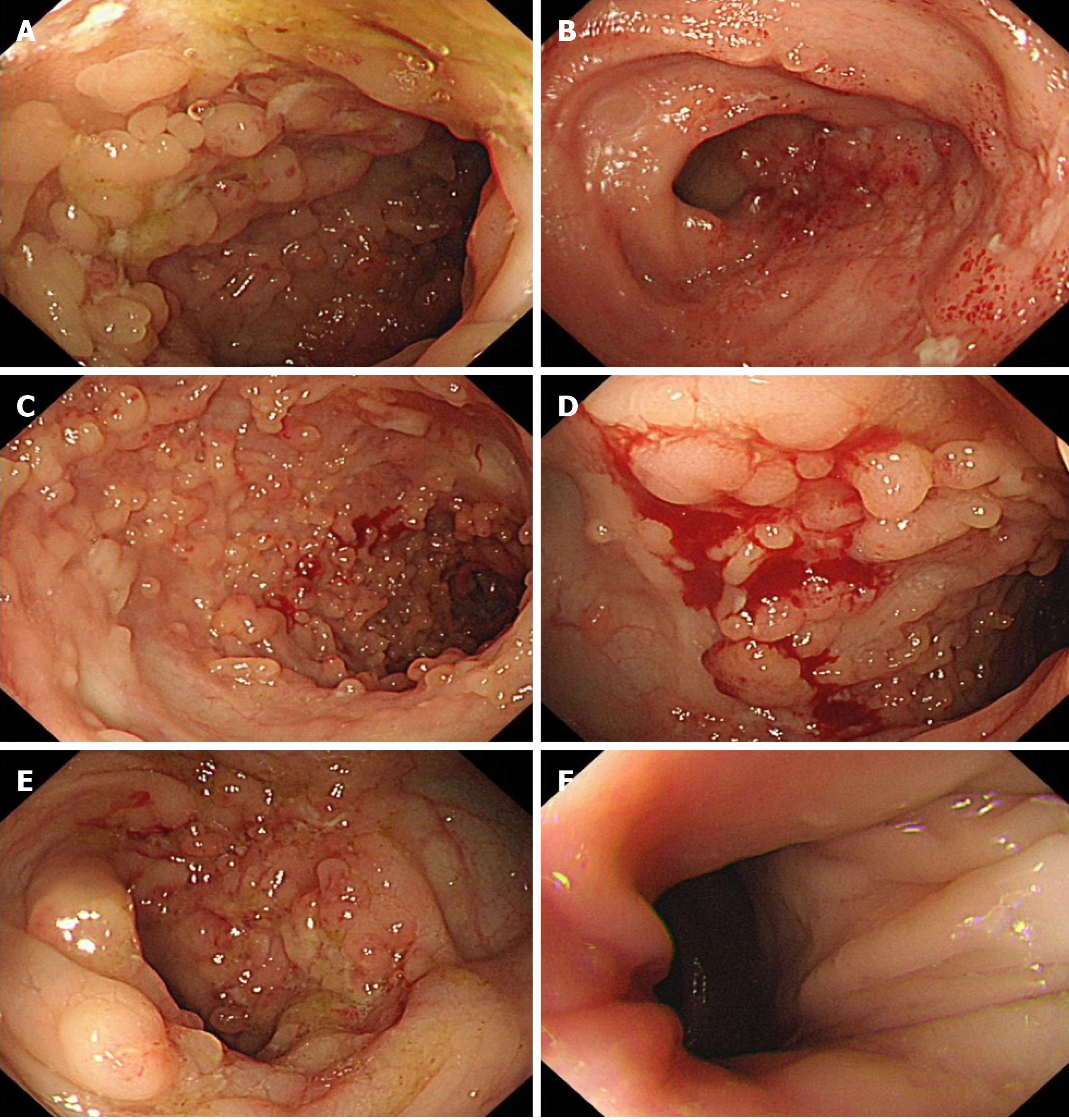
Figure 3 Colonoscopy results.
A: Distal ileum; B: Distal ileum; C: Distal ileum; D: Surgical repair site; E: Hepatic flexure; F: Anus. The distal ileal mucosa is congested and edematous, with visible erosion and scattered patchy ulcers, covered with white fur and distributed in segments. The surrounding mucosa is accompanied by pseudopolypoid hyperplasia, presenting as cobblestone-like changes. Local mucosal protrusions, ulcers, and nodular protrusions can be seen near the hepatic flexure of the transverse colon. Suspected formation of a sinus in the anus.
- Citation: Tang YJ, Zhang J, Wang J, Tian RD, Zhong WW, Yao BS, Hou BY, Chen YH, He W, He YH. Link between mutations in ACVRL1 and PLA2G4A genes and chronic intestinal ulcers: A case report and review of literature. World J Gastrointest Surg 2024; 16(3): 932-943
- URL: https://www.wjgnet.com/1948-9366/full/v16/i3/932.htm
- DOI: https://dx.doi.org/10.4240/wjgs.v16.i3.932


