Copyright
©The Author(s) 2024.
World J Gastrointest Surg. Mar 27, 2024; 16(3): 932-943
Published online Mar 27, 2024. doi: 10.4240/wjgs.v16.i3.932
Published online Mar 27, 2024. doi: 10.4240/wjgs.v16.i3.932
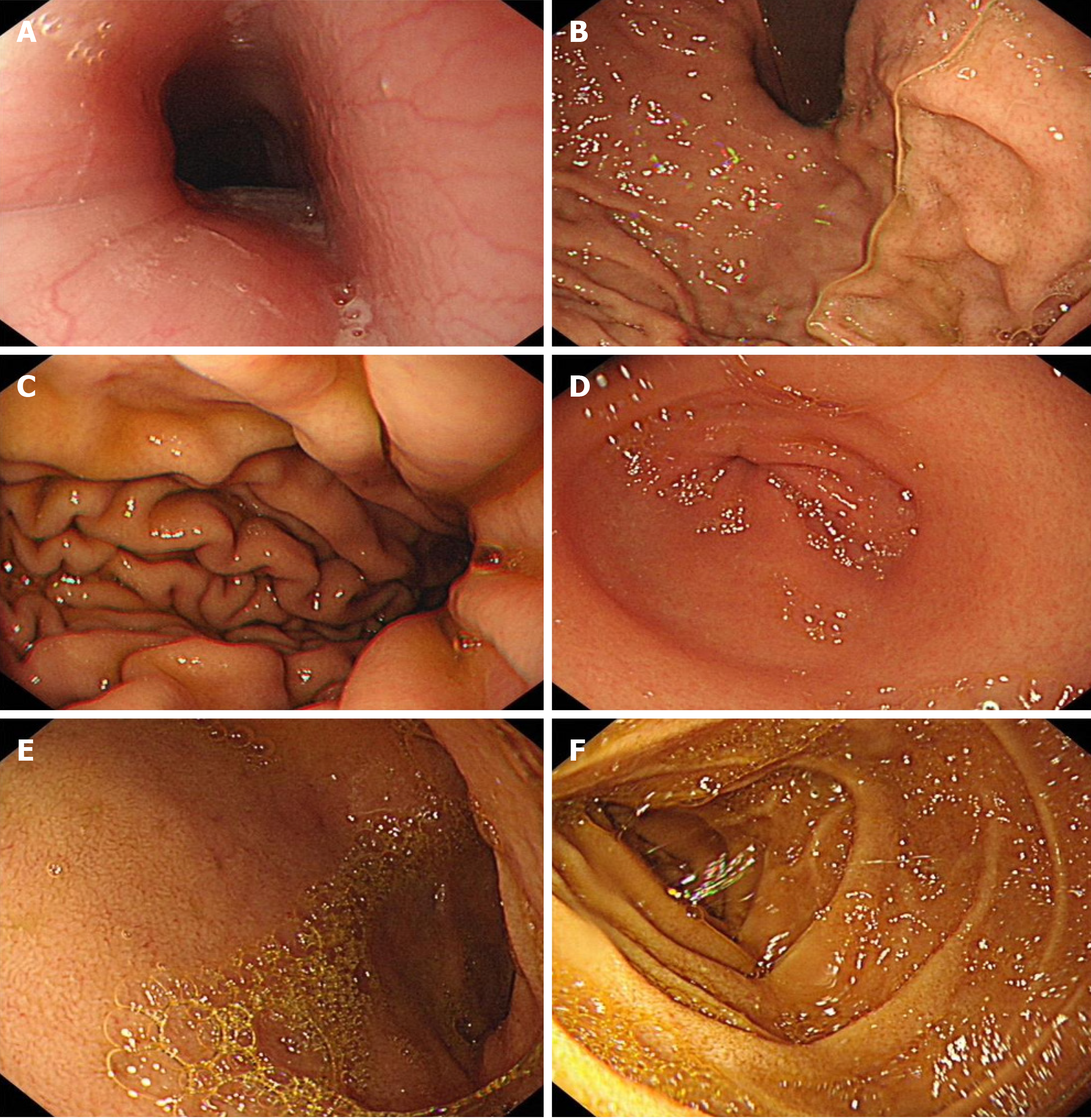
Figure 2 Gastroscopy results.
A: Esophagus; B: Gastric fundus; C: Gastric body; D: Gastric antrum; E: Duodenal bulb; F: Descending duodenum. No obvious abnormalities are seen in the morphology and color of the esophagus. The distance between the cardia and the incisor is about 40 cm, and the dentate line is clear. The gastric fundus does not show any obvious abnormalities in the mucosa and morphology, and there is a moderate amount of mucus and yellow turbidity. The gastric body's mucosa and morphology are also normal; the gastric angle is curved and smooth. The gastric antrum's mucosa is congested and edematous, but no ulcers or masses are present. The pylorus is circular in shape and can open and close smoothly. There are no obvious abnormalities in the duodenal bulb and descending mucosa.
- Citation: Tang YJ, Zhang J, Wang J, Tian RD, Zhong WW, Yao BS, Hou BY, Chen YH, He W, He YH. Link between mutations in ACVRL1 and PLA2G4A genes and chronic intestinal ulcers: A case report and review of literature. World J Gastrointest Surg 2024; 16(3): 932-943
- URL: https://www.wjgnet.com/1948-9366/full/v16/i3/932.htm
- DOI: https://dx.doi.org/10.4240/wjgs.v16.i3.932


