Copyright
©The Author(s) 2024.
World J Gastrointest Surg. Feb 27, 2024; 16(2): 601-608
Published online Feb 27, 2024. doi: 10.4240/wjgs.v16.i2.601
Published online Feb 27, 2024. doi: 10.4240/wjgs.v16.i2.601
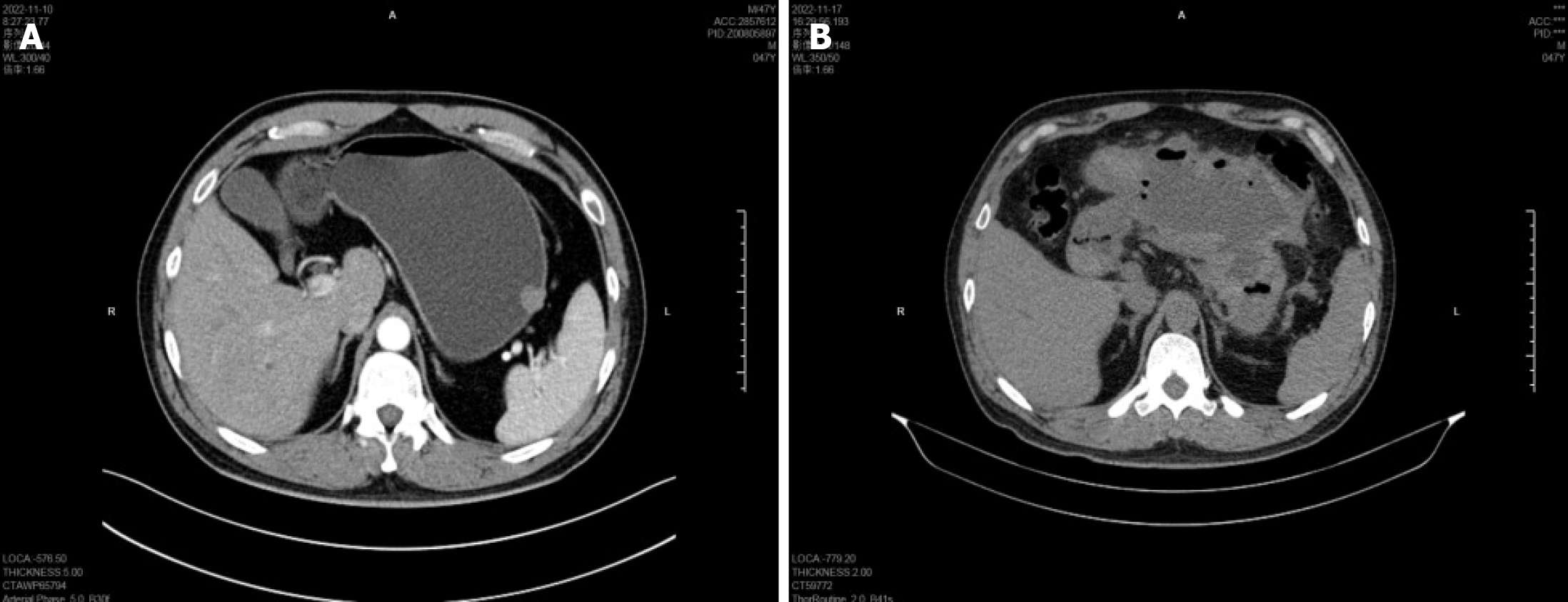
Figure 2 Abdominal computed tomography examination.
A: Gastric computed tomography (CT) illustrated a well-demarcated mass, 16 mm × 15 mm in size, demonstrating mild enhancement, with an intact mucosal line; B: Abdominal CT revealed a large amount of encapsulated fluid and gas accumulation around the stomach.
- Citation: Lu HF, Li JJ, Zhu DB, Mao LQ, Xu LF, Yu J, Yao LH. Postoperative encapsulated hemoperitoneum in a patient with gastric stromal tumor treated by exposed endoscopic full-thickness resection: A case report. World J Gastrointest Surg 2024; 16(2): 601-608
- URL: https://www.wjgnet.com/1948-9366/full/v16/i2/601.htm
- DOI: https://dx.doi.org/10.4240/wjgs.v16.i2.601


