Copyright
©The Author(s) 2024.
World J Gastrointest Surg. Dec 27, 2024; 16(12): 3806-3817
Published online Dec 27, 2024. doi: 10.4240/wjgs.v16.i12.3806
Published online Dec 27, 2024. doi: 10.4240/wjgs.v16.i12.3806
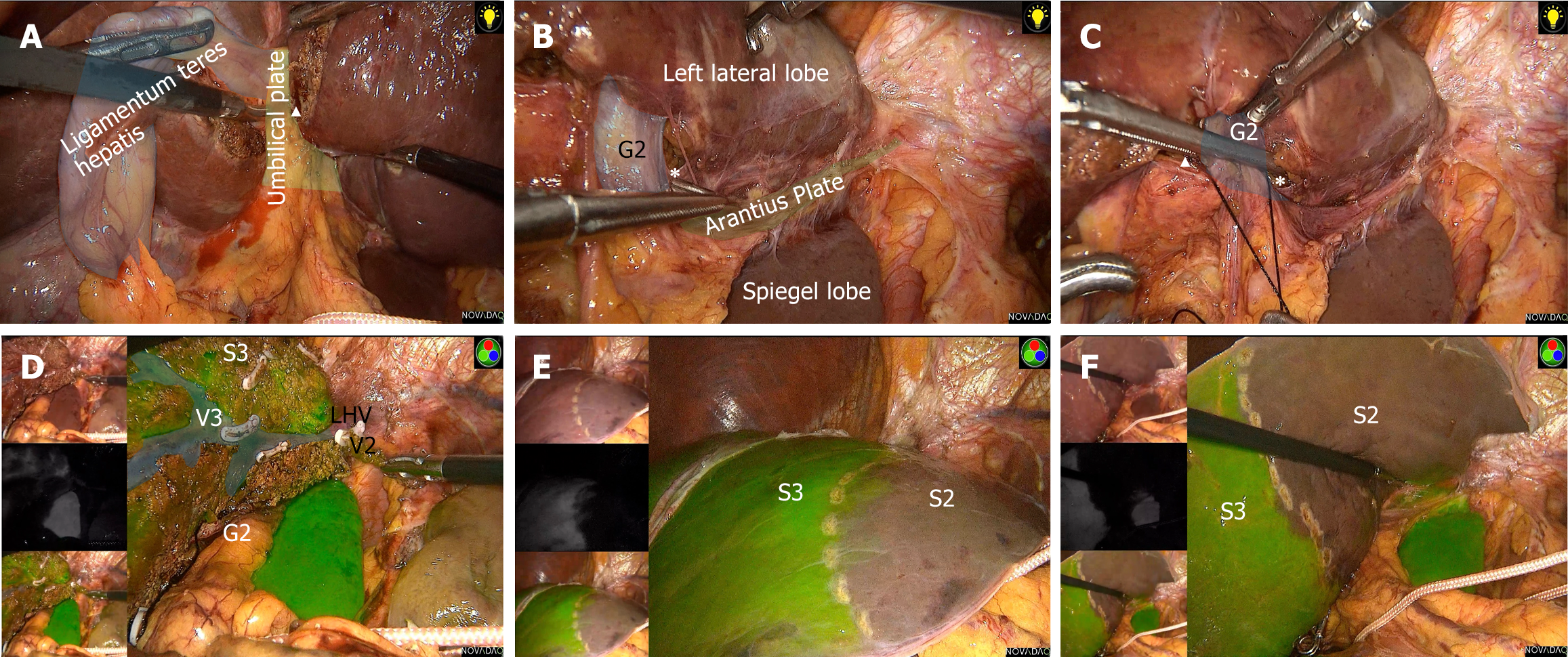
Figure 2 Segmentectomy 2.
A: Beginning from the umbilical plate; B: Exposing the caudal end of the Arantius plate; C: Dissecting the Glissonean pedicle of segment 2; D: Liver parenchyma resection along the markings; E and F: Indocyanine green staining. G2: Glissonean pedicle of segment 2; S2: Segment 2; S3: Segment 3; V3: Hepatic vein of segment 3; LHV: Left hepatic vein.
- Citation: Wang SD, Wang L, Xiao H, Chen K, Liu JR, Chen Z, Lan X. Novel techniques of liver segmental and subsegmental pedicle anatomy from segment 1 to segment 8. World J Gastrointest Surg 2024; 16(12): 3806-3817
- URL: https://www.wjgnet.com/1948-9366/full/v16/i12/3806.htm
- DOI: https://dx.doi.org/10.4240/wjgs.v16.i12.3806


