Copyright
©The Author(s) 2024.
World J Gastrointest Surg. Nov 27, 2024; 16(11): 3598-3605
Published online Nov 27, 2024. doi: 10.4240/wjgs.v16.i11.3598
Published online Nov 27, 2024. doi: 10.4240/wjgs.v16.i11.3598
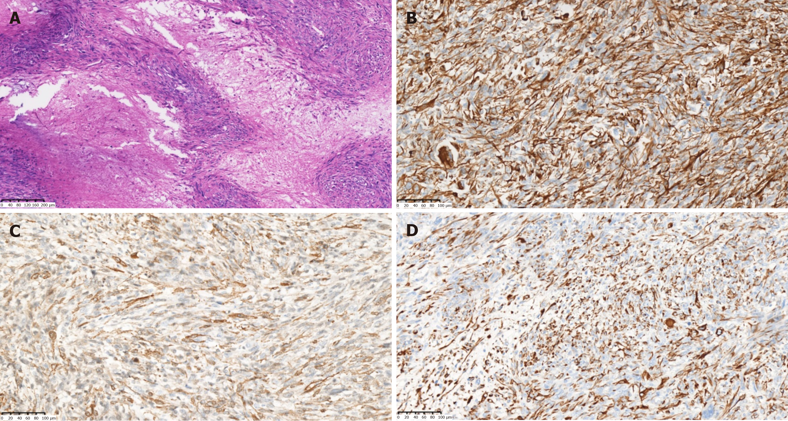
Figure 5 Postoperative pathology.
A: Large areas of map-like necrosis were found in the tumor. The tumor cells were spindle-shaped and in a bunch-like arrangement, with obvious cell atypia, varying sizes, and frequent mitotic images (hematoxylin and eosin staining, × 100); B: Vimentin (+) (hematoxylin and eosin staining, × 200); C: Anti-smooth muscle actin (+) (immunohistochemical staining, × 200); D: Desmin (+) (immunohistochemical staining, × 200).
- Citation: Wu FN, Zhang M, Zhang K, Lv XL, Guo JQ, Tu CY, Zhou QY. Primary hepatic leiomyosarcoma masquerading as liver abscess: A case report. World J Gastrointest Surg 2024; 16(11): 3598-3605
- URL: https://www.wjgnet.com/1948-9366/full/v16/i11/3598.htm
- DOI: https://dx.doi.org/10.4240/wjgs.v16.i11.3598


