Copyright
©The Author(s) 2024.
World J Gastrointest Surg. Nov 27, 2024; 16(11): 3598-3605
Published online Nov 27, 2024. doi: 10.4240/wjgs.v16.i11.3598
Published online Nov 27, 2024. doi: 10.4240/wjgs.v16.i11.3598
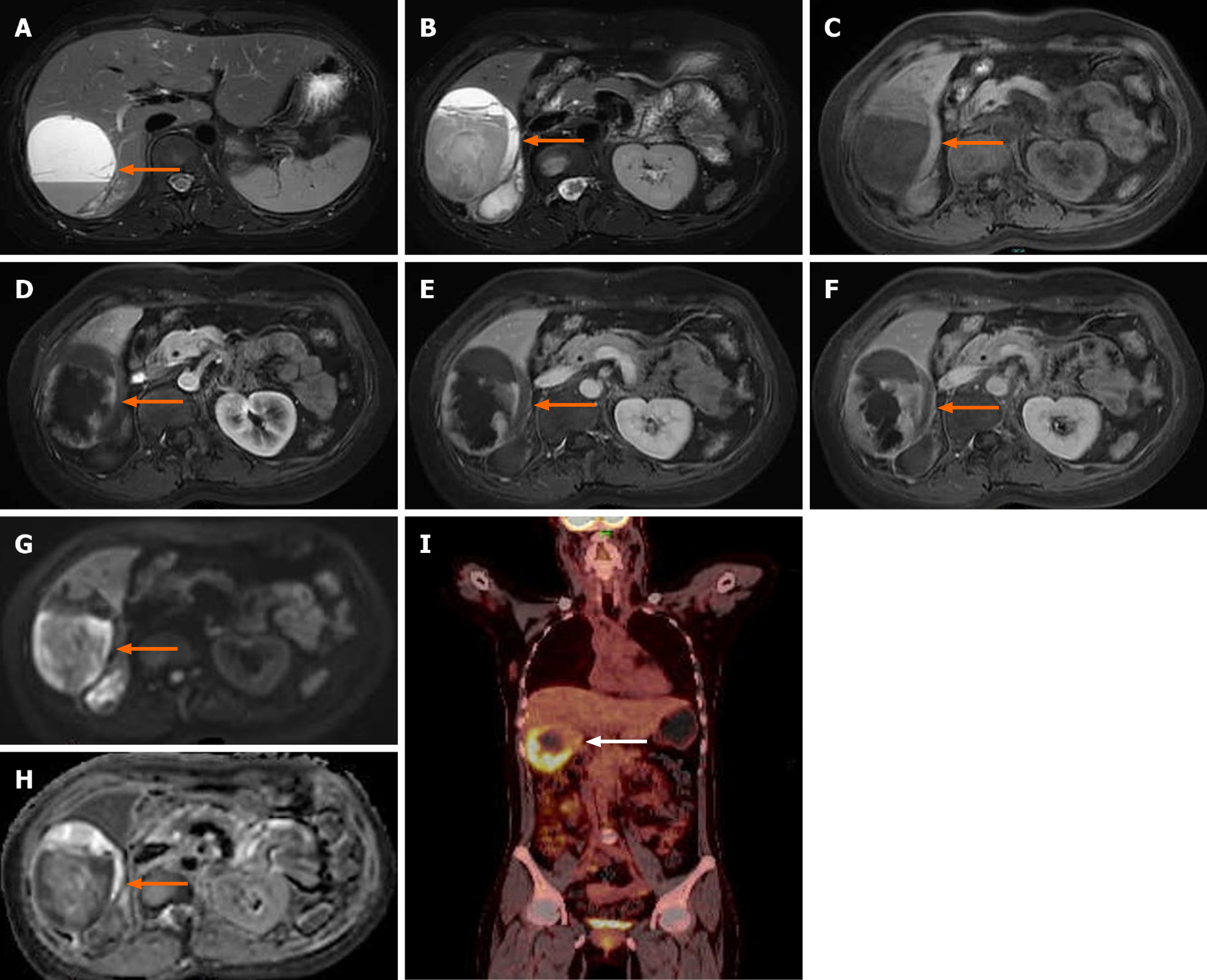
Figure 2 Magnetic resonance imaging of the upper abdomen and positron emission tomography-computed tomography.
Magnetic resonance imaging showed a massive abnormal signal lesion in the right lobe of the liver, with septations and fluid levels. The lesion showed heterogeneous hypointensity on T1-weighted imaging and heterogeneous hyperintensity on T2-weighted imaging. The edges and septations of the lesion were significantly enhanced during the arterial phase, and the enhancement partially fused and filled toward the center of the lesion during the portal phase and delayed phase. Diffusion-weighted imaging showed a hyperintense signal and the corresponding apparent diffusion coefficient map showed a decreased signal intensity in parts of the wall of the lesion (the arrow shows the lesion). The positron emission tomography-computed tomography scan showed an irregular low-density lesion in the right lobe of the liver with increased uptake (the arrow shows the lesion). A and B: T2-weighted imaging; C: T1-weighted imaging; D: Diffusion-weighted imaging; E: Apparent diffusion coefficient; F: The arterial phase of the enhanced scan; G: The portal vein phase; H: Delayed phase; I: Positron emission tomography-computed tomography.
- Citation: Wu FN, Zhang M, Zhang K, Lv XL, Guo JQ, Tu CY, Zhou QY. Primary hepatic leiomyosarcoma masquerading as liver abscess: A case report. World J Gastrointest Surg 2024; 16(11): 3598-3605
- URL: https://www.wjgnet.com/1948-9366/full/v16/i11/3598.htm
- DOI: https://dx.doi.org/10.4240/wjgs.v16.i11.3598


