Copyright
©The Author(s) 2024.
World J Gastrointest Surg. Nov 27, 2024; 16(11): 3598-3605
Published online Nov 27, 2024. doi: 10.4240/wjgs.v16.i11.3598
Published online Nov 27, 2024. doi: 10.4240/wjgs.v16.i11.3598
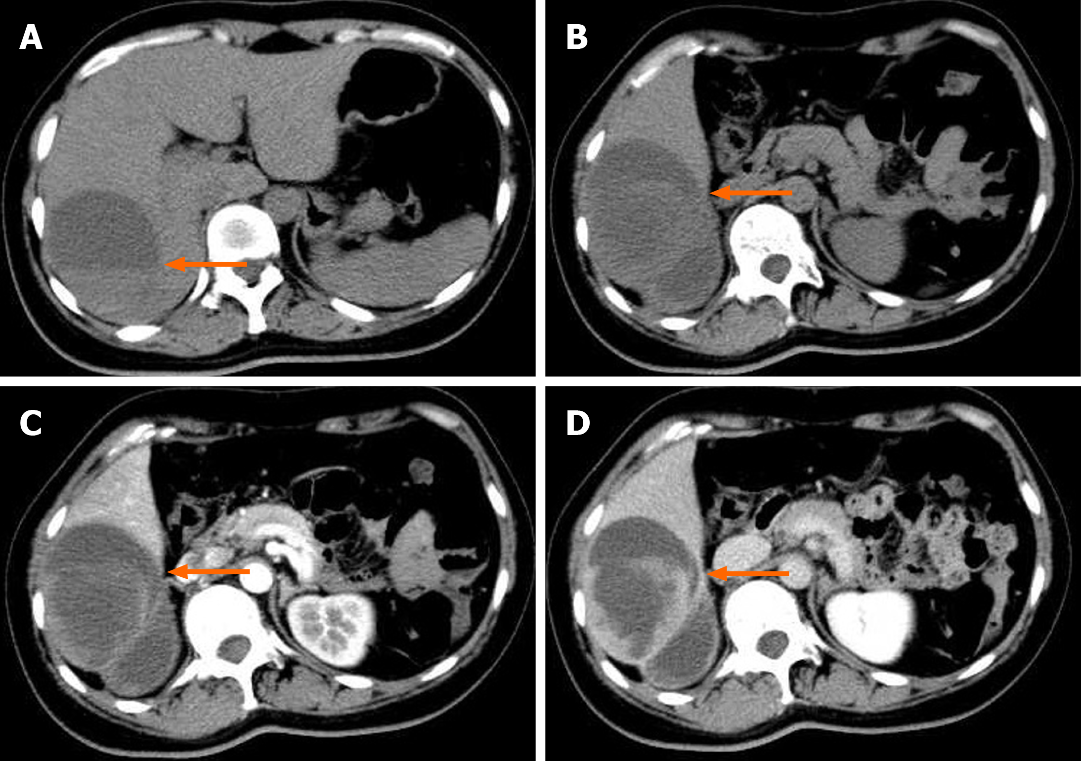
Figure 1 Computed tomography of the upper abdomen before surgery.
A mixed low-density lesion (6.5 cm × 9.7 cm × 9.6 cm) can be seen in the right lobe of the liver, within which septations and fluid levels are visible. The enhanced scan showed significant enhancement of the lesion margin and septation (the arrow shows the lesion). A: Plain computed tomography scan; B: Plain computed tomography scan; C: The arterial phase of the enhanced scan; D: Delayed phase.
- Citation: Wu FN, Zhang M, Zhang K, Lv XL, Guo JQ, Tu CY, Zhou QY. Primary hepatic leiomyosarcoma masquerading as liver abscess: A case report. World J Gastrointest Surg 2024; 16(11): 3598-3605
- URL: https://www.wjgnet.com/1948-9366/full/v16/i11/3598.htm
- DOI: https://dx.doi.org/10.4240/wjgs.v16.i11.3598


