Copyright
©The Author(s) 2024.
World J Gastrointest Surg. Nov 27, 2024; 16(11): 3590-3597
Published online Nov 27, 2024. doi: 10.4240/wjgs.v16.i11.3590
Published online Nov 27, 2024. doi: 10.4240/wjgs.v16.i11.3590
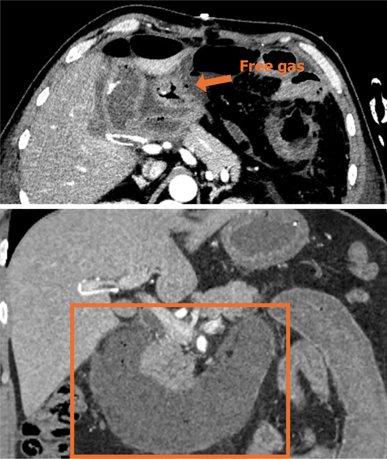
Figure 1 Preoperative contrast-enhanced computed tomography of the abdominopelvic cavity.
Diffuse thickening of the gastric wall and duodenal intestinal wall in the gastric sinus department, blurring of the plasma membrane surface, blurring of the peripheral fat interstitial space with multiple plaques and cord shadows, a few gas-dense shadows were seen at the edge of the gastric sinus, and the head of the neighboring pancreas was also seen. The head of the neighboring pancreas was full, with blurred borders, the dilatation of the lumen of the gastric sinus and the duodenal tube, and the edema and thickening of the wall of the gastric sinus. Thickening of the wall of the proximal jejunum. Diagnostic imaging considered sulcus pancreatitis suspected perforation of the gastric sinus.
- Citation: Tong KN, Zhang WT, Liu K, Xu R, Guo W. Emergency pancreaticoduodenectomy for pancreatitis-associated necrotic perforation of the distal stomach and full-length duodenum: A case report. World J Gastrointest Surg 2024; 16(11): 3590-3597
- URL: https://www.wjgnet.com/1948-9366/full/v16/i11/3590.htm
- DOI: https://dx.doi.org/10.4240/wjgs.v16.i11.3590


