Copyright
©The Author(s) 2024.
World J Gastrointest Surg. Nov 27, 2024; 16(11): 3584-3589
Published online Nov 27, 2024. doi: 10.4240/wjgs.v16.i11.3584
Published online Nov 27, 2024. doi: 10.4240/wjgs.v16.i11.3584
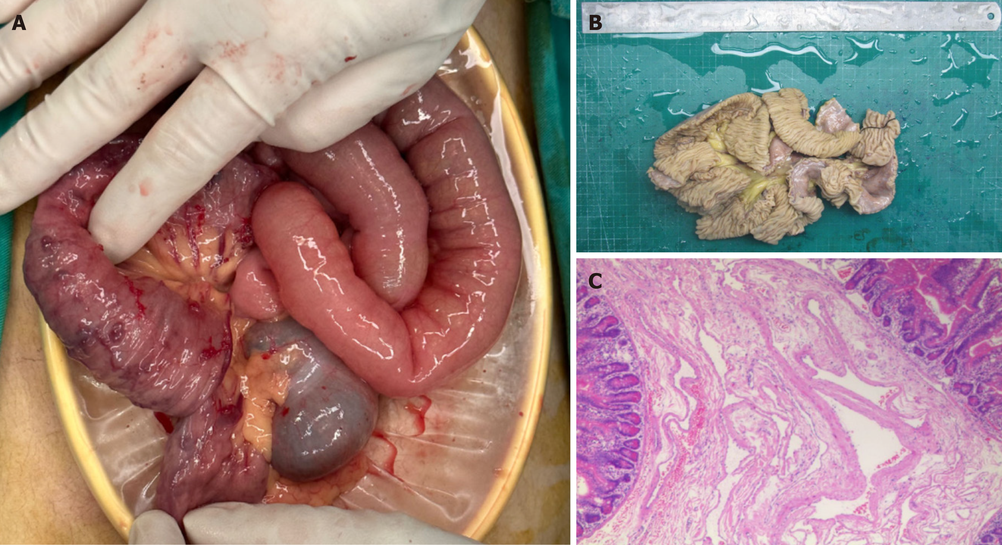
Figure 2 Intestinal lesions.
A: Diseased small intestine (left) and normal small intestine (right); B: Diseased small intestine after resection; C: Multiple venous malformations under the small intestinal mucosa.
- Citation: Wang WJ, Chen PL, Shao HZ. Blue rubber blister nevus syndrome: A case report. World J Gastrointest Surg 2024; 16(11): 3584-3589
- URL: https://www.wjgnet.com/1948-9366/full/v16/i11/3584.htm
- DOI: https://dx.doi.org/10.4240/wjgs.v16.i11.3584


