Copyright
©The Author(s) 2024.
World J Gastrointest Surg. Oct 27, 2024; 16(10): 3350-3357
Published online Oct 27, 2024. doi: 10.4240/wjgs.v16.i10.3350
Published online Oct 27, 2024. doi: 10.4240/wjgs.v16.i10.3350
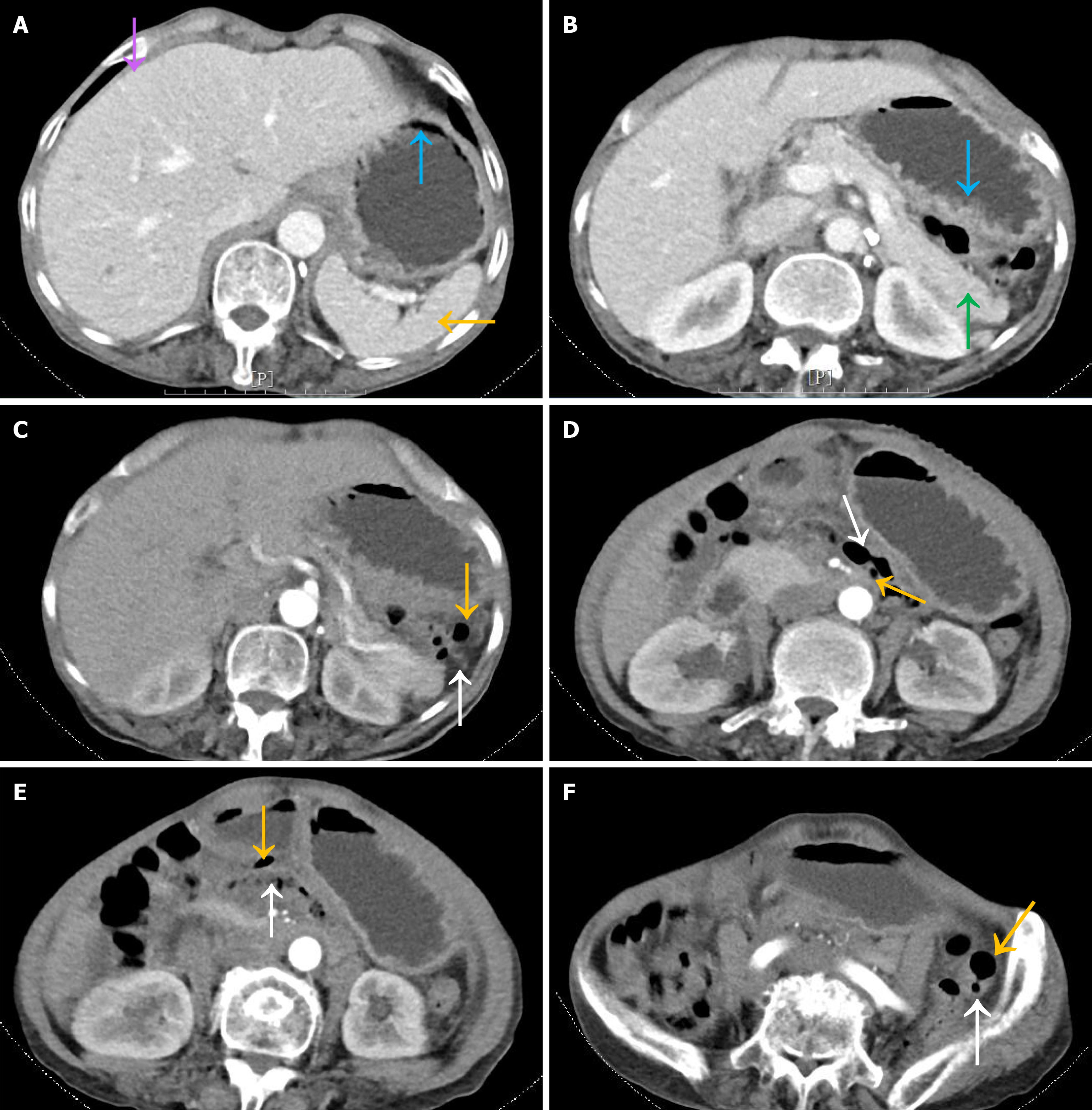
Figure 5 Abdominal enhanced computed tomography examination.
A: Shows the disappearance of free air in the abdominal cavity (purple arrows), improvement of gastric dilatation, normal gastric morphology (blue arrows), and return of the spleen to a normal anatomical position (orange arrows); B: Shows no thickened mass in the gastric wall (blue arrows) and normal pancreatic morphology (green arrows); C-F: Multiple cases of subserosal pneumatosis in the colon (orange arrows) with intact continuity of the bowel wall (white arrows).
- Citation: Zhang Q, Xu XJ, Ma J, Huang HY, Zhang YM. Acute gastric volvulus combined with pneumatosis coli rupture misdiagnosed as gastric volvulus with perforation: A case report. World J Gastrointest Surg 2024; 16(10): 3350-3357
- URL: https://www.wjgnet.com/1948-9366/full/v16/i10/3350.htm
- DOI: https://dx.doi.org/10.4240/wjgs.v16.i10.3350


