Copyright
©The Author(s) 2024.
World J Gastrointest Surg. Oct 27, 2024; 16(10): 3350-3357
Published online Oct 27, 2024. doi: 10.4240/wjgs.v16.i10.3350
Published online Oct 27, 2024. doi: 10.4240/wjgs.v16.i10.3350
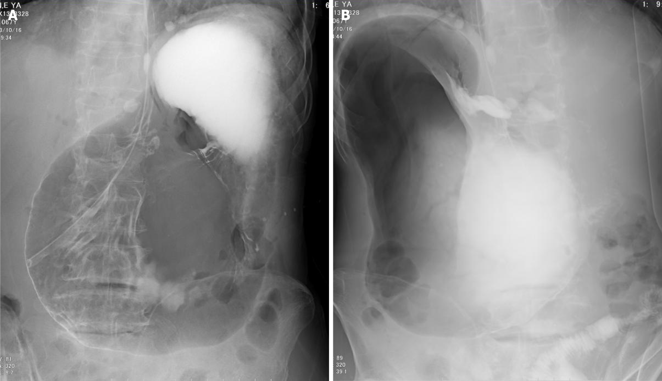
Figure 3 Upper gastrointestinal imaging examination upon admission.
A: Shows the anterior view of upper gastrointestinal radiography, the contrast agent is limited, and the gastric lumen is not completely visualized; B: The posterior view of upper gastrointestinal radiography, the gastric lumen is dilated with pneumatosis, and the greater and lesser curvature sides cannot be distinguished.
- Citation: Zhang Q, Xu XJ, Ma J, Huang HY, Zhang YM. Acute gastric volvulus combined with pneumatosis coli rupture misdiagnosed as gastric volvulus with perforation: A case report. World J Gastrointest Surg 2024; 16(10): 3350-3357
- URL: https://www.wjgnet.com/1948-9366/full/v16/i10/3350.htm
- DOI: https://dx.doi.org/10.4240/wjgs.v16.i10.3350


