Copyright
©The Author(s) 2024.
World J Gastrointest Surg. Oct 27, 2024; 16(10): 3350-3357
Published online Oct 27, 2024. doi: 10.4240/wjgs.v16.i10.3350
Published online Oct 27, 2024. doi: 10.4240/wjgs.v16.i10.3350
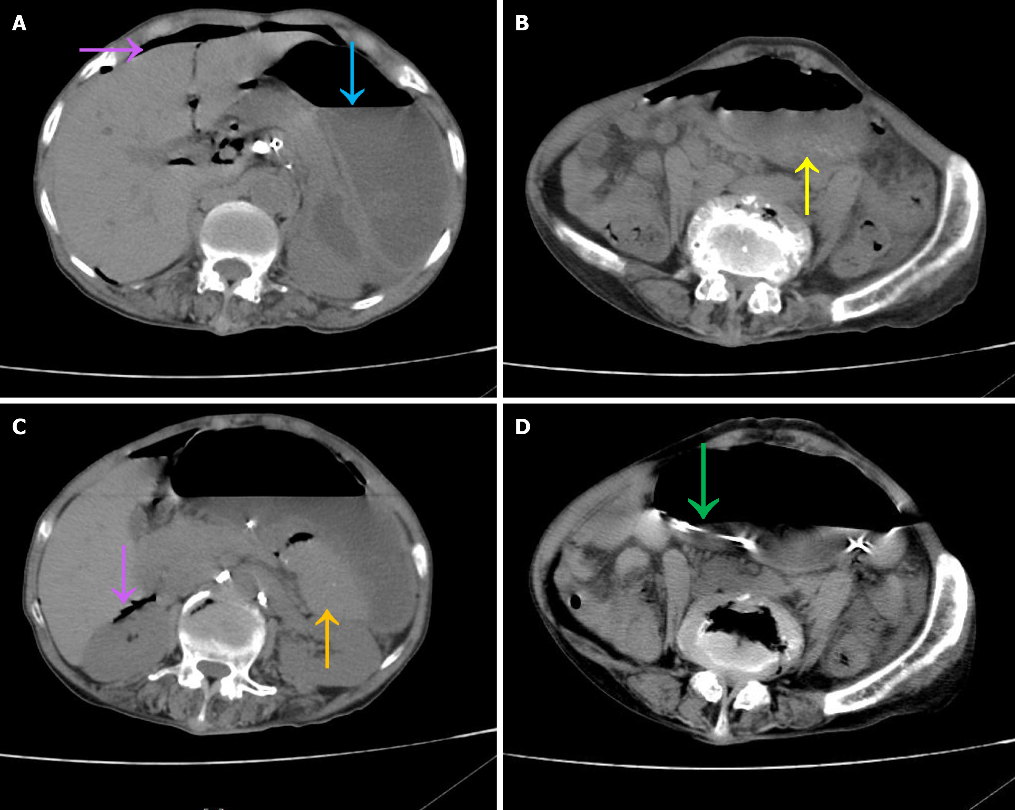
Figure 1 Abdominal computed tomography scan upon admission.
A: Free intraperitoneal air without fluid collection (purple arrow) and a markedly dilated gastric lumen with fluid collection (blue arrow); B: The lower gastric margin reaching the level of the anterior superior iliac spine (yellow arrow); C: Spleen displacement to the upper pancreatic margin (orange arrow) and free intraperitoneal air (purple arrow); D: The gastric tube entering the gastric body from left to right through the posterior aspect of the stomach (green arrow).
- Citation: Zhang Q, Xu XJ, Ma J, Huang HY, Zhang YM. Acute gastric volvulus combined with pneumatosis coli rupture misdiagnosed as gastric volvulus with perforation: A case report. World J Gastrointest Surg 2024; 16(10): 3350-3357
- URL: https://www.wjgnet.com/1948-9366/full/v16/i10/3350.htm
- DOI: https://dx.doi.org/10.4240/wjgs.v16.i10.3350


