Copyright
©The Author(s) 2024.
World J Gastrointest Surg. Oct 27, 2024; 16(10): 3343-3349
Published online Oct 27, 2024. doi: 10.4240/wjgs.v16.i10.3343
Published online Oct 27, 2024. doi: 10.4240/wjgs.v16.i10.3343
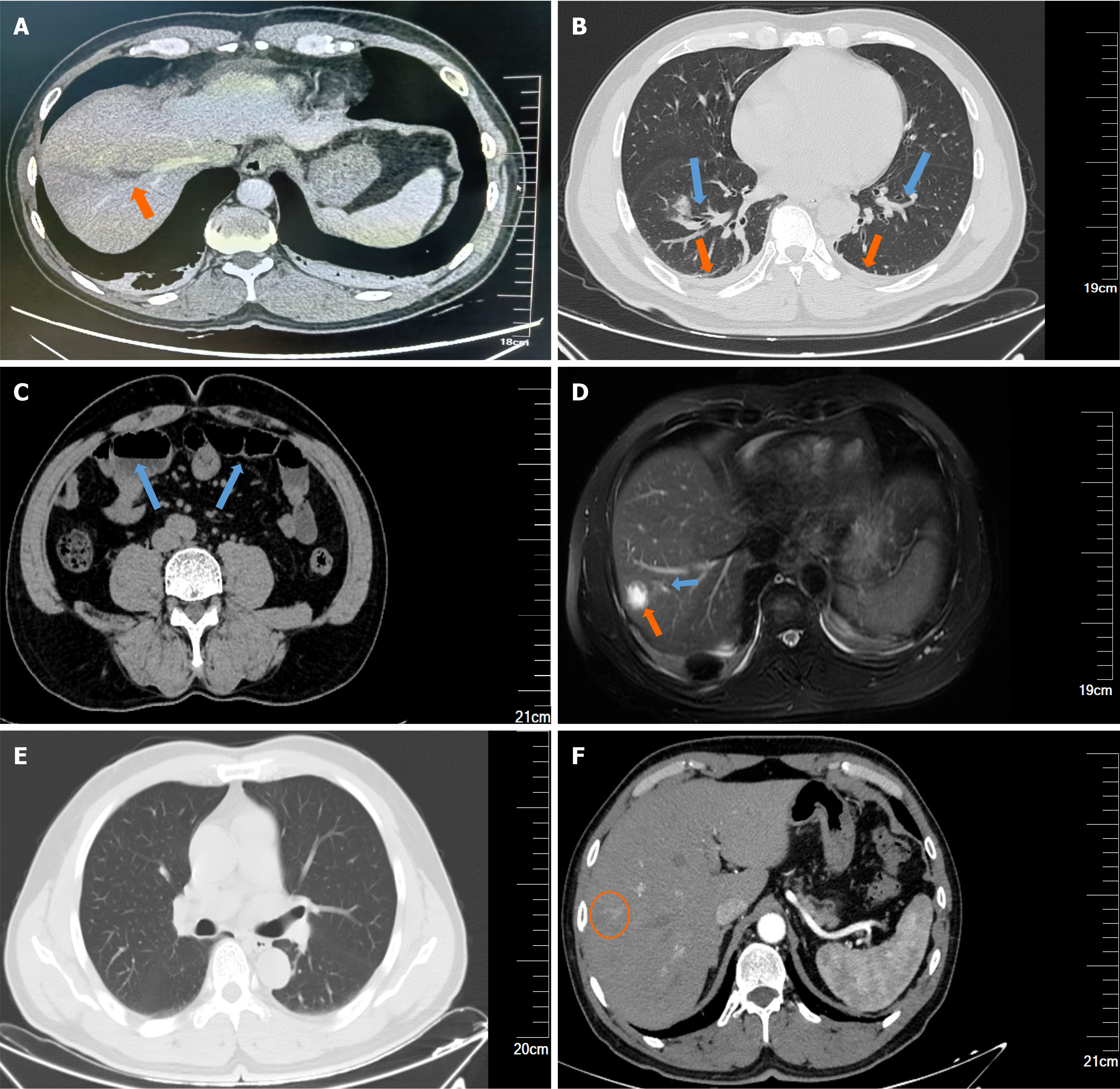
Figure 1 Imaging examination results of the patient.
A: Hepatic enhanced computed tomography (CT) showed hepatic thrombophlebitis in the right liver lobe (orange arrow: Filling defect in the vein with irregular wall thickness); B: Abdominal CT showed bilateral lung inflammation (blue arrow) and bilateral pleural effusion (orange arrow); C: Intestinal CT showed fluid accumulation in the small intestine and bowel dilatation (blue arrow). During a 56-day follow-up; D: Magnetic resonance cholangiopancreatography showed a right liver lobe abscess and purulent cholangitis (orange arrow: 20 mm × 16 mm liver abscess; blue arrow: Purulent cholangitis with thickened duct walls); E: Abdominal CT showed absorbed pneumonia and pleural effusion; F: Hepatic enhanced CT revealed low-density nodule (approximately 6 mm) in the upper segment of the right lobe (S8) indicating granulomatous changes after liver abscess treatment (orange circle) and complete resolution of the hepatic vein thrombus.
- Citation: Niu CY, Yao BT, Tao HY, Peng XG, Zhang QH, Chen Y, Liu L. Leukopenia-a rare complication secondary to invasive liver abscess syndrome in a patient with diabetes mellitus: A case report. World J Gastrointest Surg 2024; 16(10): 3343-3349
- URL: https://www.wjgnet.com/1948-9366/full/v16/i10/3343.htm
- DOI: https://dx.doi.org/10.4240/wjgs.v16.i10.3343


