Copyright
©The Author(s) 2024.
World J Gastrointest Surg. Oct 27, 2024; 16(10): 3334-3342
Published online Oct 27, 2024. doi: 10.4240/wjgs.v16.i10.3334
Published online Oct 27, 2024. doi: 10.4240/wjgs.v16.i10.3334
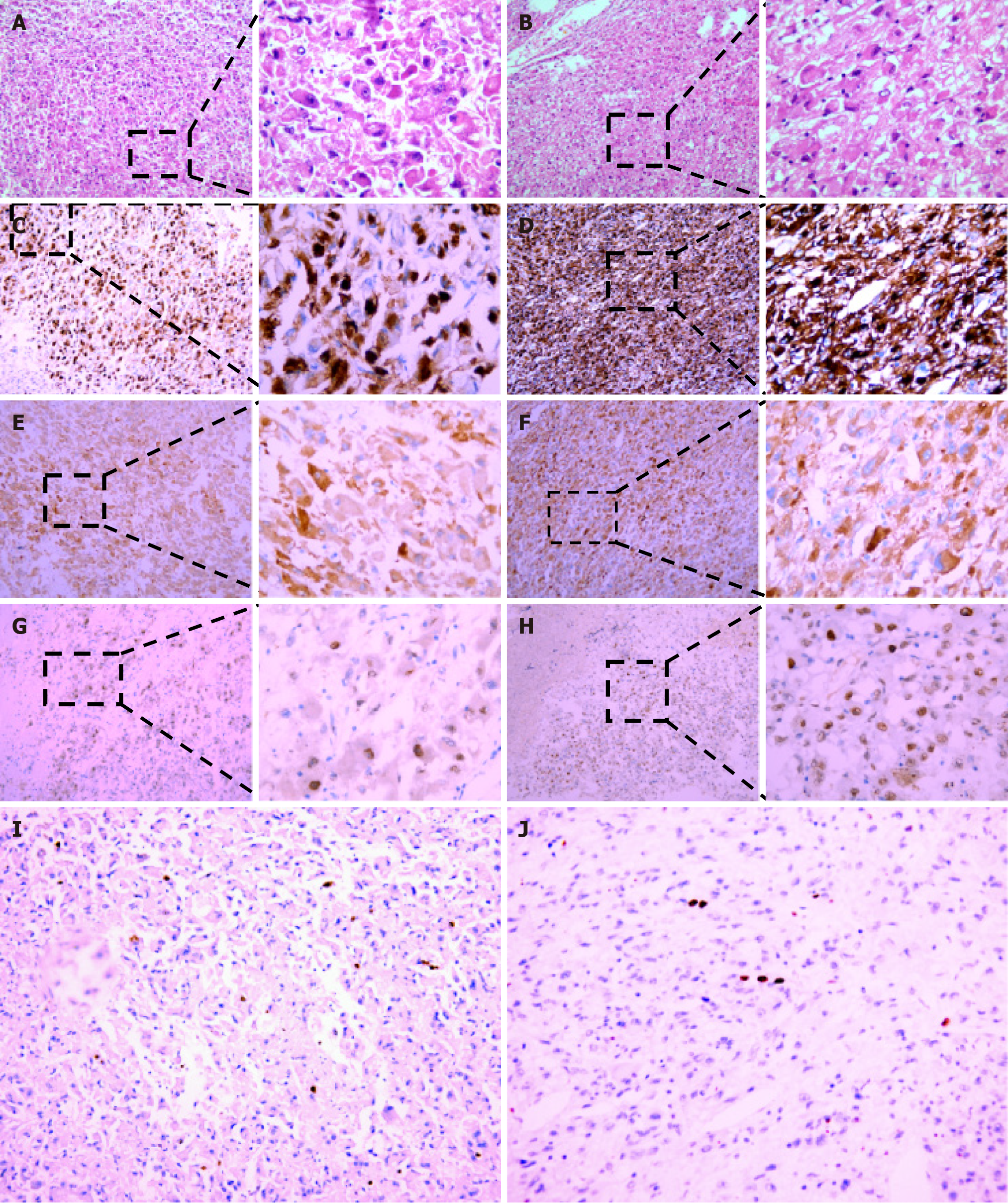
Figure 6 Pathological findings of surgical specimens.
A and B: The histopathological examination of surgical specimens from liver and kidney lesions showed tumor lesions mainly composed of dysplasia epithelioid cells with rich and red-stained cytoplasm and large, atypical and vacuolated nuclei, and prominent nucleolus (hematoxylin-eosin, original magnification × 100 and × 400); C and D: The surgical specimens from the liver and kidney lesions showed strong and diffuse granular cytoplasmic immunoreactivity for HMB-45 in epithelioid tumor cells (original magnification × 100 and × 400); E and F: The surgical specimens from the liver and kidney lesions exhibited intense and partly cytoplasmic expression of melan-A, with a staining pattern similar to that of HMB-45 (original magnification × 100 and × 400); G and H: The surgical specimens from the liver and kidney lesions revealed weak and punctate nuclear immunoreactivity for TFE3 in the epithelioid tumor cells (original magnification × 100 and × 400); I and J: The surgical specimens from the liver and kidney lesions displayed weak expression of Ki67 in the nuclei of the epithelioid cells (original magnification × 100).
- Citation: Yang HT, Wang FR, He N, She YH, Du YY, Shi WG, Yang J, Chen G, Zhang SZ, Cui F, Long B, Yu ZY, Zhu JM, Zhang GY. Massive simultaneous hepatic and renal perivascular epithelioid cell tumor benefitted from surgery and everolimus treatment: A case report. World J Gastrointest Surg 2024; 16(10): 3334-3342
- URL: https://www.wjgnet.com/1948-9366/full/v16/i10/3334.htm
- DOI: https://dx.doi.org/10.4240/wjgs.v16.i10.3334


