Copyright
©The Author(s) 2024.
World J Gastrointest Surg. Oct 27, 2024; 16(10): 3334-3342
Published online Oct 27, 2024. doi: 10.4240/wjgs.v16.i10.3334
Published online Oct 27, 2024. doi: 10.4240/wjgs.v16.i10.3334
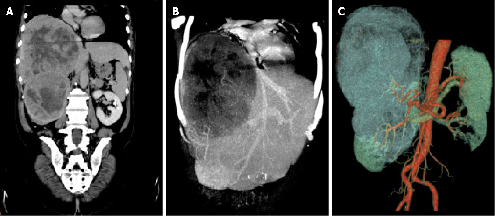
Figure 3 Images of tumor vascular supply.
A: The computed tomography reconstructions displayed a large well-circumscribed mass with uneven density in the right lobe of the liver and a clear-boundary and round-like neoplasm in the right kidney at the coronal level; B: In the maximum intensity projection images, there was no visualization of the right hepatic vein and the middle hepatic vein appeared compressed and deformed due to the presence of the tumor; C: The volume rendering technique images revealed that the hepatic tumor was supplied by branches of the proper hepatic artery and the renal neoplasm received blood supply from branches of the renal artery.
- Citation: Yang HT, Wang FR, He N, She YH, Du YY, Shi WG, Yang J, Chen G, Zhang SZ, Cui F, Long B, Yu ZY, Zhu JM, Zhang GY. Massive simultaneous hepatic and renal perivascular epithelioid cell tumor benefitted from surgery and everolimus treatment: A case report. World J Gastrointest Surg 2024; 16(10): 3334-3342
- URL: https://www.wjgnet.com/1948-9366/full/v16/i10/3334.htm
- DOI: https://dx.doi.org/10.4240/wjgs.v16.i10.3334


