Copyright
©The Author(s) 2024.
World J Gastrointest Surg. Oct 27, 2024; 16(10): 3321-3327
Published online Oct 27, 2024. doi: 10.4240/wjgs.v16.i10.3321
Published online Oct 27, 2024. doi: 10.4240/wjgs.v16.i10.3321
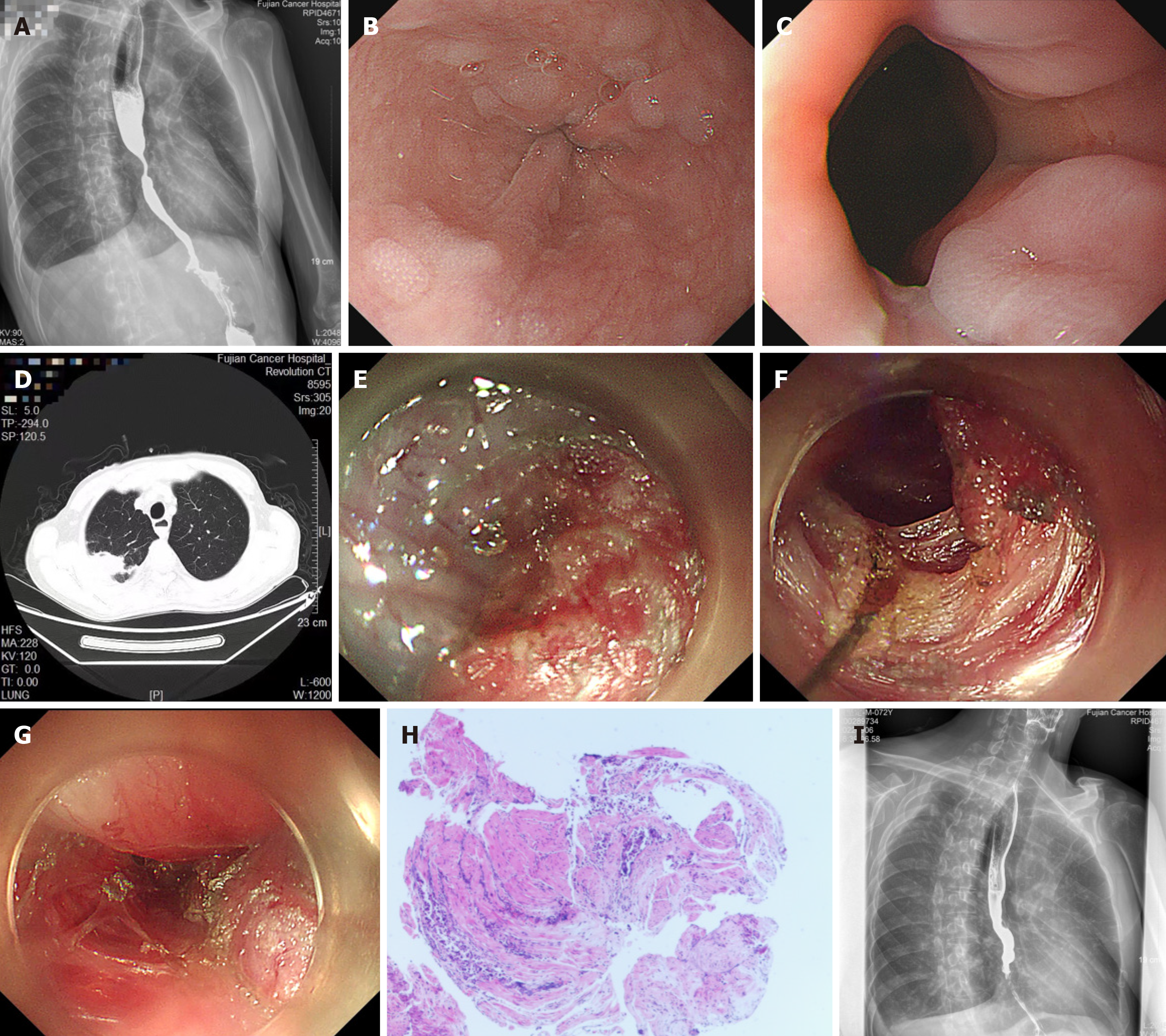
Figure 2 Diagnosis and treatment of the patient.
A: Esophagogram revealed grade I dilation of the esophageal lumen, segmental stenosis of the distal esophagus, and delayed emptying of barium contrast agent from the esophagus; B: Endoscopy revealed a 2-cm long segment of concentric esophageal stenosis with apparently normal overlying mucosa; C: The cardia had normal contraction and relaxation; D: Computed tomography revealed a right upper lung mass; E: Unclear hierarchy of the esophageal wall at the narrow segment; F: Myotomy at the narrow segment; G: The length of the entire myotomy was approximately 6 cm; H: Esophageal muscle biopsy result; I: Repeated esophagography on postoperative day 54.
- Citation: Shi H, Chen SY, Xie ZF, Lin LL, Jiang Y. Lung cancer metastasis-induced distal esophageal segmental spasm confirmed by individualized peroral endoscopic myotomy: A case report. World J Gastrointest Surg 2024; 16(10): 3321-3327
- URL: https://www.wjgnet.com/1948-9366/full/v16/i10/3321.htm
- DOI: https://dx.doi.org/10.4240/wjgs.v16.i10.3321


