Copyright
©The Author(s) 2024.
World J Gastrointest Surg. Jan 27, 2024; 16(1): 13-20
Published online Jan 27, 2024. doi: 10.4240/wjgs.v16.i1.13
Published online Jan 27, 2024. doi: 10.4240/wjgs.v16.i1.13
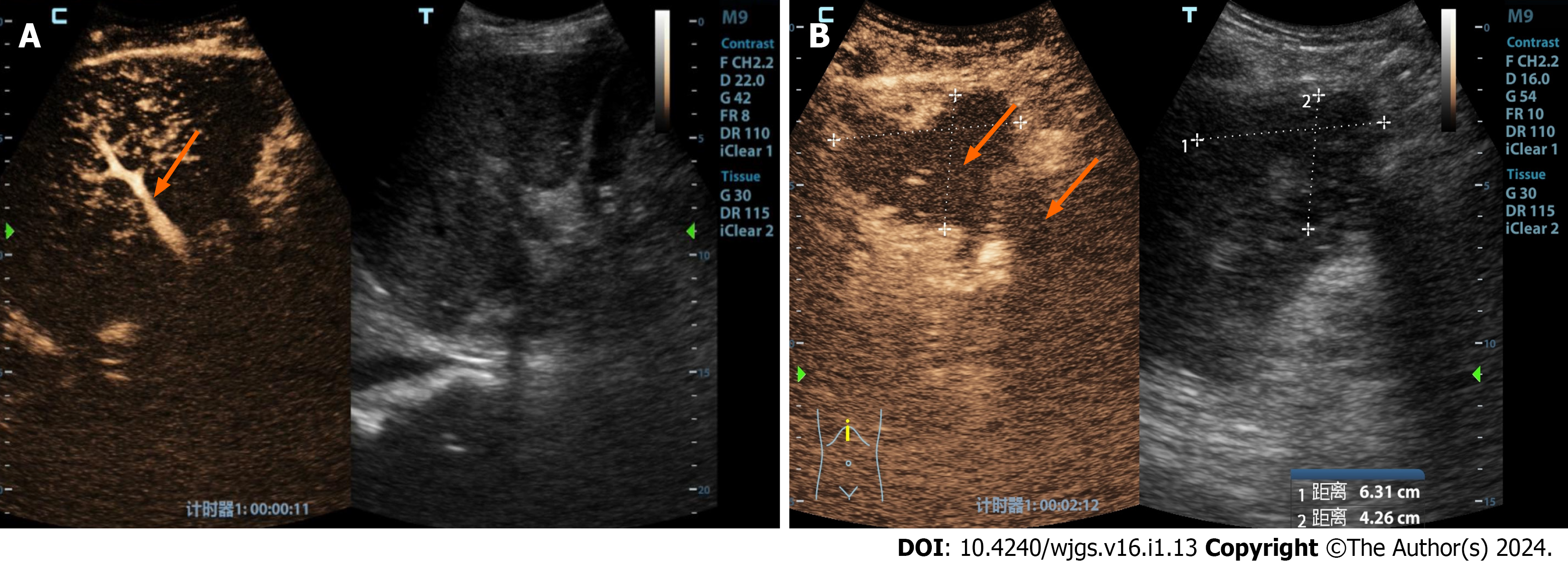
Figure 1 A 32-year-old male patient underwent a piggyback liver transplant for decompensated cirrhosis and acute liver failure.
A: This assessment occurred 15 d postoperatively. Routine ultrasound imaging had failed to reveal hepatic arteries within both the liver and extrahepatic regions. Contrast-enhanced ultrasound showed that the portal vein was prematurely visible (indicated by the orange arrow), while neither intrahepatic nor extrahepatic arteries displayed any enhancement; B: Furthermore, multiple ischemic lesions within the liver were observed in the arterial (phase I), portal (phase II), and late phases (phase III), indicating a lack of enhancement (as indicated by the orange arrow). Surgical confirmation subsequently identified a diffuse formation of arterial thrombosis within the hepatic artery.
- Citation: Zhao NB, Chen Y, Xia R, Tang JB, Zhao D. Prognostic value of ultrasound in early arterial complications post liver transplant. World J Gastrointest Surg 2024; 16(1): 13-20
- URL: https://www.wjgnet.com/1948-9366/full/v16/i1/13.htm
- DOI: https://dx.doi.org/10.4240/wjgs.v16.i1.13


