Copyright
©The Author(s) 2023.
World J Gastrointest Surg. Sep 27, 2023; 15(9): 2042-2051
Published online Sep 27, 2023. doi: 10.4240/wjgs.v15.i9.2042
Published online Sep 27, 2023. doi: 10.4240/wjgs.v15.i9.2042
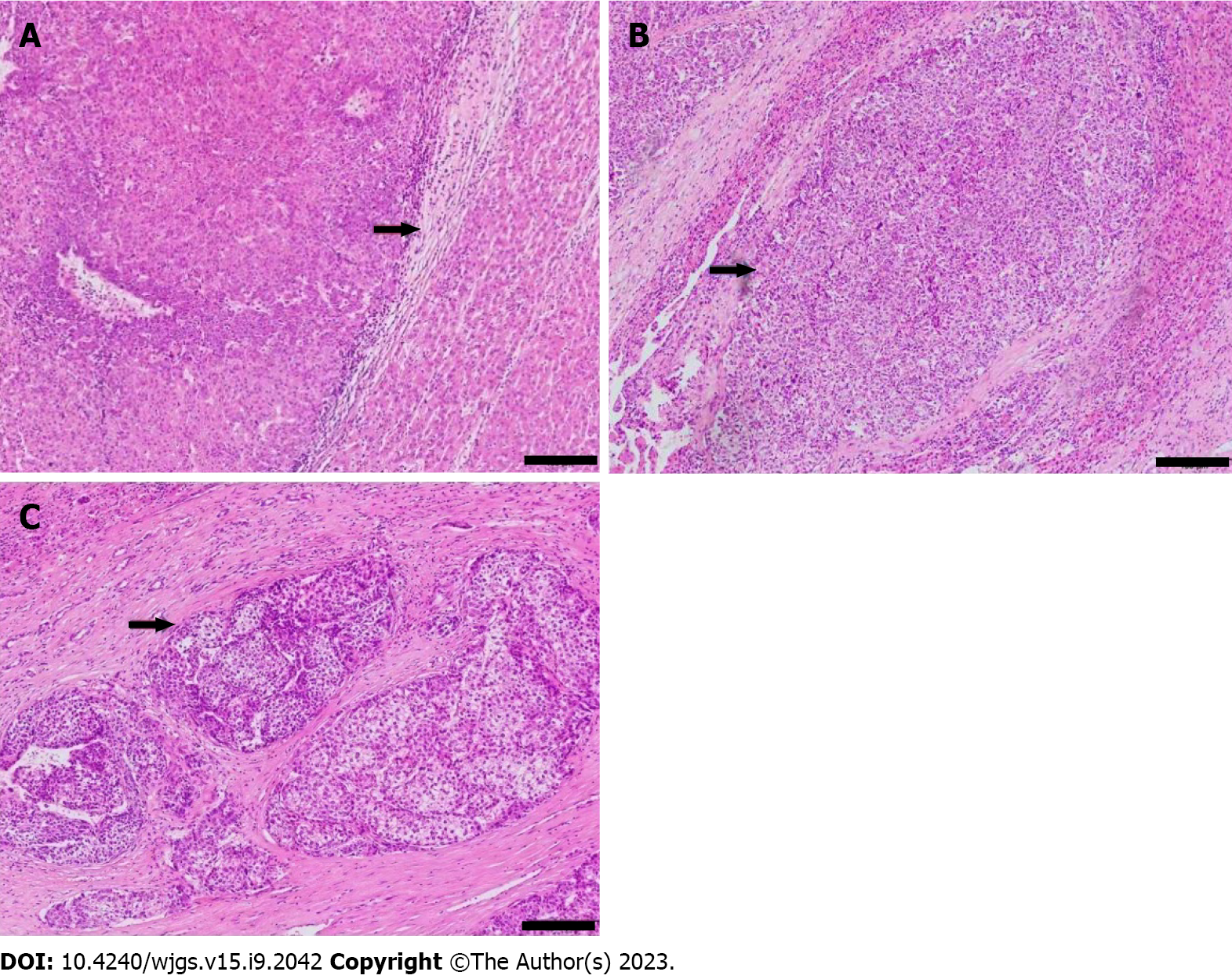
Figure 3 Pathological images showed microvascular invasion in hepatocellular carcinoma.
Hematoxylin and eosin stain, magnification: × 100. A: No microvascular invasion (MVI) detected, recorded as MVI-negative; B: One MVI detected, recorded as mild MVI; C: More than 5 MVIs detected, recorded as severe MVI. Scale bar: 100 μm.
- Citation: Jiang D, Qian Y, Tan BB, Zhu XL, Dong H, Qian R. Preoperative prediction of microvascular invasion in hepatocellular carcinoma using ultrasound features including elasticity. World J Gastrointest Surg 2023; 15(9): 2042-2051
- URL: https://www.wjgnet.com/1948-9366/full/v15/i9/2042.htm
- DOI: https://dx.doi.org/10.4240/wjgs.v15.i9.2042


