Copyright
©The Author(s) 2023.
World J Gastrointest Surg. Sep 27, 2023; 15(9): 1841-1857
Published online Sep 27, 2023. doi: 10.4240/wjgs.v15.i9.1841
Published online Sep 27, 2023. doi: 10.4240/wjgs.v15.i9.1841
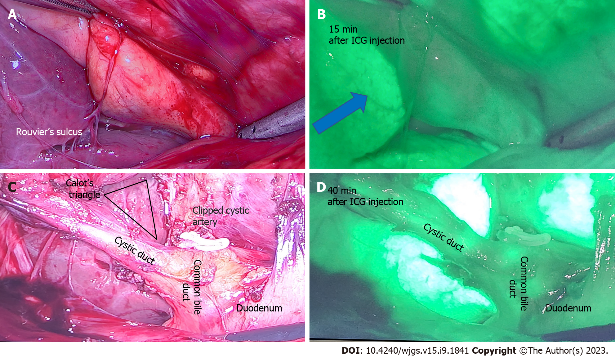
Figure 4 A 50-year-old patient undergoing elective laparoscopic cholecystectomy for previous acute cholecystitis was injected with 4 mL of indocyanine green dye after insertion of camera port.
A: Rouvier’s sulcus and corresponding; B: After 15 min of injection shows the dye enhances the liver (blue arrow) and indocyanine green (ICG) is yet to be excreted in biliary tree; C: Calot’s triangle with a critical view of safety and clipped cystic artery; D: At 40 min after ICG injection shows beginning of biliary excretion in cystic duct and common bile duct. ICG: Indocyanine green.
- Citation: Lim ZY, Mohan S, Balasubramaniam S, Ahmed S, Siew CCH, Shelat VG. Indocyanine green dye and its application in gastrointestinal surgery: The future is bright green. World J Gastrointest Surg 2023; 15(9): 1841-1857
- URL: https://www.wjgnet.com/1948-9366/full/v15/i9/1841.htm
- DOI: https://dx.doi.org/10.4240/wjgs.v15.i9.1841


