Copyright
©The Author(s) 2023.
World J Gastrointest Surg. Aug 27, 2023; 15(8): 1574-1590
Published online Aug 27, 2023. doi: 10.4240/wjgs.v15.i8.1574
Published online Aug 27, 2023. doi: 10.4240/wjgs.v15.i8.1574
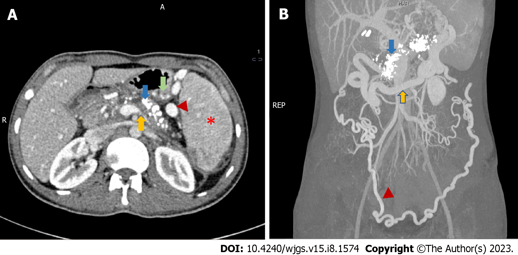
Figure 2 Radiological features of chronic calcific pancreatitis and its complications including venous thrombosis and collaterals.
A: An axial section of contrast-enhanced computed tomography (CECT) showing chronic pancreatitis with calcifications (blue arrow), attenuated splenic vein (yellow arrow), multiple perigastric collaterals (green arrow), gastrosplenic collaterals (red arrowhead) and splenomegaly (red asterisk); B: Coronal section of CECT of the same patient showing extensive pancreatic calcification (blue arrow) with dilated gastroepiploic vein (yellow arrow) and omental collaterals (red arrowhead). Courtesy: Dr Madhusudhan KS, Department of Radiodiagnosis.
- Citation: Walia D, Saraya A, Gunjan D. Vascular complications of chronic pancreatitis and its management. World J Gastrointest Surg 2023; 15(8): 1574-1590
- URL: https://www.wjgnet.com/1948-9366/full/v15/i8/1574.htm
- DOI: https://dx.doi.org/10.4240/wjgs.v15.i8.1574


