Copyright
©The Author(s) 2023.
World J Gastrointest Surg. Jun 27, 2023; 15(6): 1240-1246
Published online Jun 27, 2023. doi: 10.4240/wjgs.v15.i6.1240
Published online Jun 27, 2023. doi: 10.4240/wjgs.v15.i6.1240
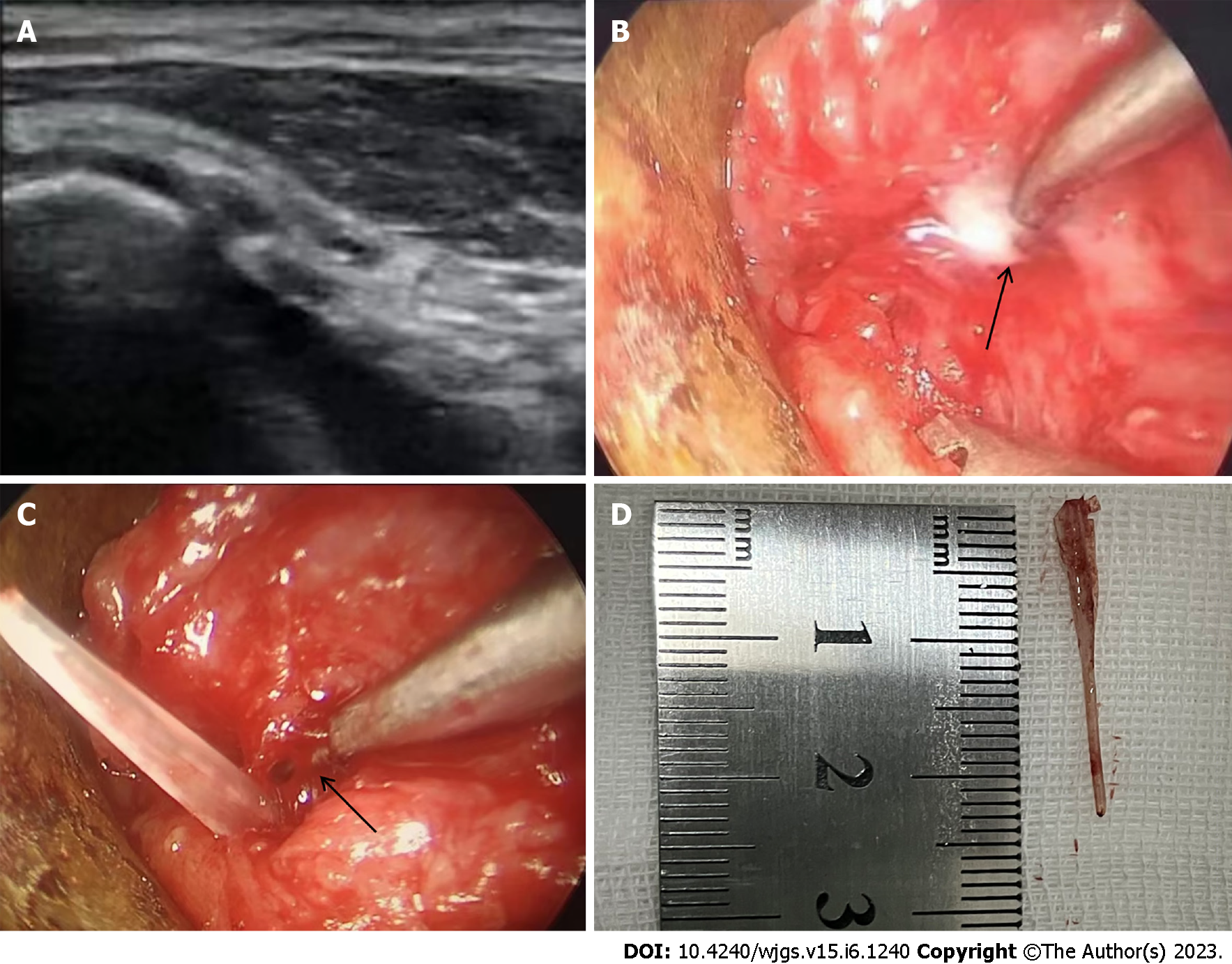
Figure 2 Operation.
A: Ultrasound imaging: The needle was punctured into the cavity where the fishbone was located; B: Endoscopy examination: A small amount of purulent fluid was seen overflowing from the dissected lateral wall of the right piriform fossa under video endoscopy and laryngoscope; C: Endoscopy examination: The head of the foreign body (fishbone) was exposed; D: Fishbone removed: The removed fish bone measured 2.5 cm in diameter.
- Citation: Wei HX, Lv SY, Xia B, Zhang K, Pan CK. Bedside ultrasound-guided water injection assists endoscopically treatment in esophageal perforation caused by foreign bodies: A case report. World J Gastrointest Surg 2023; 15(6): 1240-1246
- URL: https://www.wjgnet.com/1948-9366/full/v15/i6/1240.htm
- DOI: https://dx.doi.org/10.4240/wjgs.v15.i6.1240


