Copyright
©The Author(s) 2023.
World J Gastrointest Surg. Jun 27, 2023; 15(6): 1232-1239
Published online Jun 27, 2023. doi: 10.4240/wjgs.v15.i6.1232
Published online Jun 27, 2023. doi: 10.4240/wjgs.v15.i6.1232
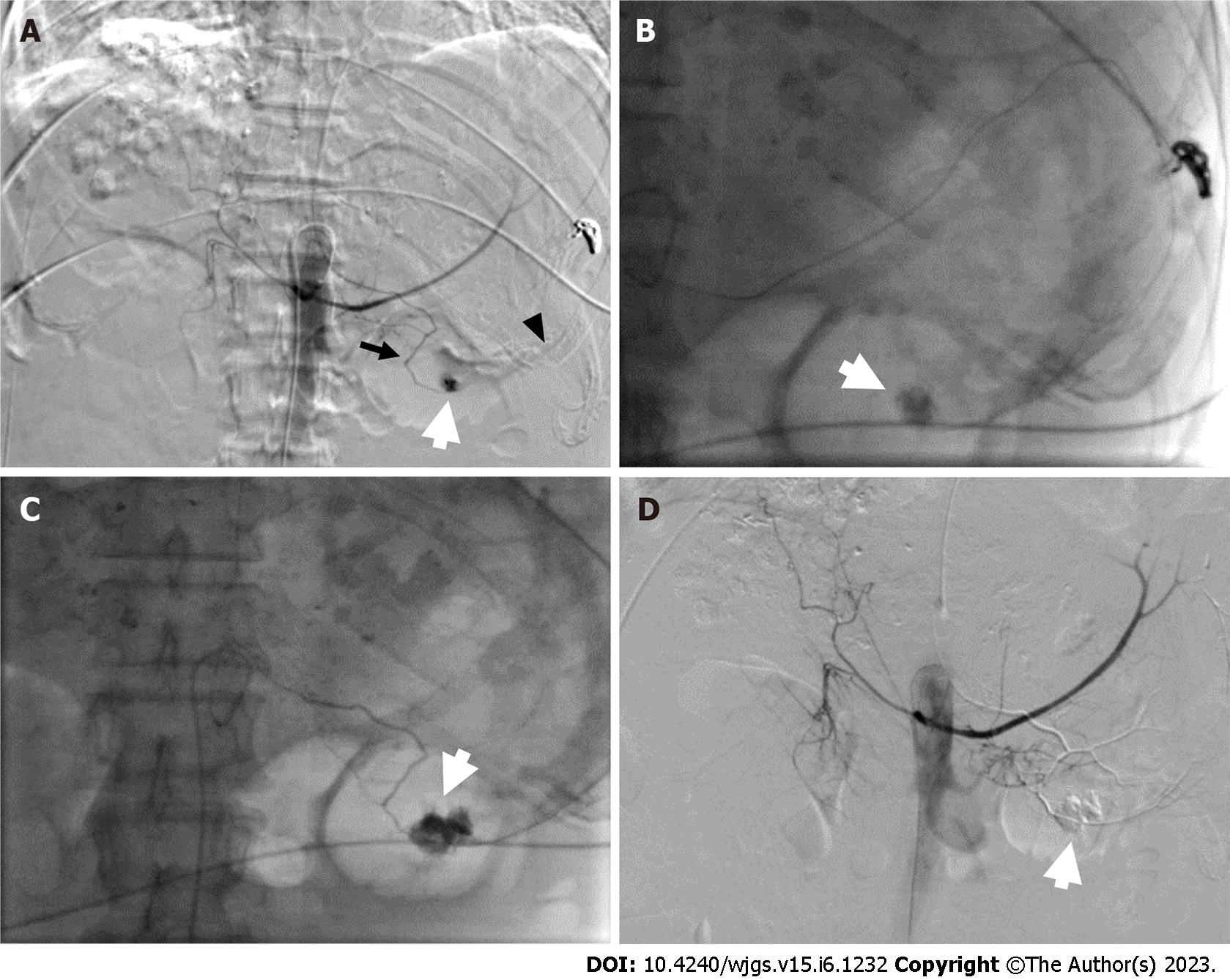
Figure 5 Emergency angiography.
A: Selective digital subtraction angiography of the celiac trunk showed extravasation of contrast medium (white arrow) from the inferior splenic artery (black arrow), a branch of the left gastric artery (black arrow head), and a pseudoaneurysm of the gastric artery; B and C: The pseudoaneurysm (white arrow) was embolized using liquid glue and lipiodol; D: After embolization, the pseudoaneurysm (white arrow) and active bleeding were no longer visible.
- Citation: Pang FW, Chen B, Peng DT, He J, Zhao WC, Chen TT, Xie ZG, Deng HH. Massive bleeding from a gastric artery pseudoaneurysm in hepatocellular carcinoma treated with atezolizumab plus bevacizumab: A case report. World J Gastrointest Surg 2023; 15(6): 1232-1239
- URL: https://www.wjgnet.com/1948-9366/full/v15/i6/1232.htm
- DOI: https://dx.doi.org/10.4240/wjgs.v15.i6.1232


