Copyright
©The Author(s) 2023.
World J Gastrointest Surg. Jun 27, 2023; 15(6): 1224-1231
Published online Jun 27, 2023. doi: 10.4240/wjgs.v15.i6.1224
Published online Jun 27, 2023. doi: 10.4240/wjgs.v15.i6.1224
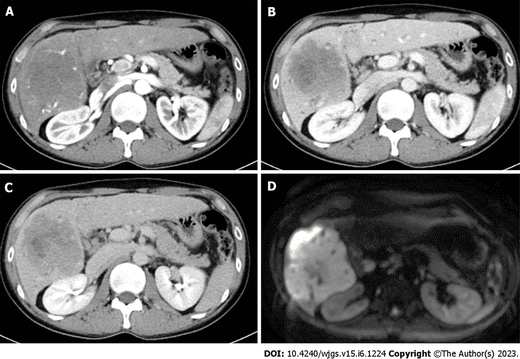
Figure 1 Abdominal contrast-enhanced computed tomography images and magnetic resonance image of case 1.
A: Early arterial phase of computed tomography (CT); B: Portal vein phase of CT; C: Late phase of CT. A massive mass with a major axis of about 10 cm almost occupies the right lobe of the liver S5-6. The mass is gradually stained in a non-uniform ring shape. D: Diffusion weighted image of magnetic resonance image.
- Citation: Miyazu T, Ishida N, Asai Y, Tamura S, Tani S, Yamade M, Iwaizumi M, Hamaya Y, Osawa S, Baba S, Sugimoto K. Intrahepatic cholangiocarcinoma in patients with primary sclerosing cholangitis and ulcerative colitis: Two case reports. World J Gastrointest Surg 2023; 15(6): 1224-1231
- URL: https://www.wjgnet.com/1948-9366/full/v15/i6/1224.htm
- DOI: https://dx.doi.org/10.4240/wjgs.v15.i6.1224


