Copyright
©The Author(s) 2023.
World J Gastrointest Surg. Jun 27, 2023; 15(6): 1216-1223
Published online Jun 27, 2023. doi: 10.4240/wjgs.v15.i6.1216
Published online Jun 27, 2023. doi: 10.4240/wjgs.v15.i6.1216
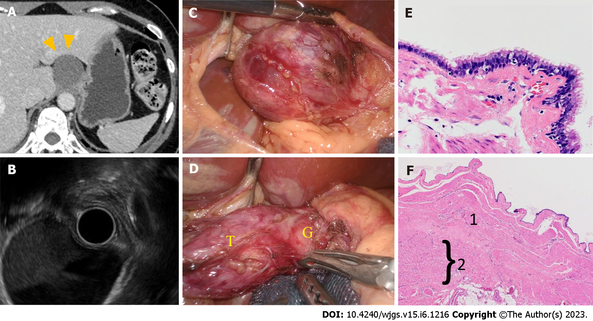
Figure 1 The findings of Patient 1.
A: Computed tomography scan of the abdomen revealed a cystic mass with a diameter of 3 cm. The lesion was attached to the gastric cardia with regular outlines (arrows); B: Endoscopic ultrasonography findings: A cystic mass was found in the submucosal layer of the cardia; C and D: Laparoscopic findings: (C) A smooth cystic mass arose from the gastric cardia and (D) part of the cyst (T) adhered firmly to the gastric wall (G); E: Histopathological findings: The cystic wall was lined with ciliated columnar epithelia and mucous glandular cells without cytological atypia; high-power magnification; F: the smooth muscle fibers surrounded the cystic wall (1) and they were continuous with the gastric muscular layer (2); low-power magnification. G: The gastric wall; T: The tumor.
- Citation: Terayama M, Kumagai K, Kawachi H, Makuuchi R, Hayami M, Ida S, Ohashi M, Sano T, Nunobe S. Optimal resection of gastric bronchogenic cysts based on anatomical continuity with adherent gastric muscular layer: A case report. World J Gastrointest Surg 2023; 15(6): 1216-1223
- URL: https://www.wjgnet.com/1948-9366/full/v15/i6/1216.htm
- DOI: https://dx.doi.org/10.4240/wjgs.v15.i6.1216


