Copyright
©The Author(s) 2023.
World J Gastrointest Surg. May 27, 2023; 15(5): 978-983
Published online May 27, 2023. doi: 10.4240/wjgs.v15.i5.978
Published online May 27, 2023. doi: 10.4240/wjgs.v15.i5.978
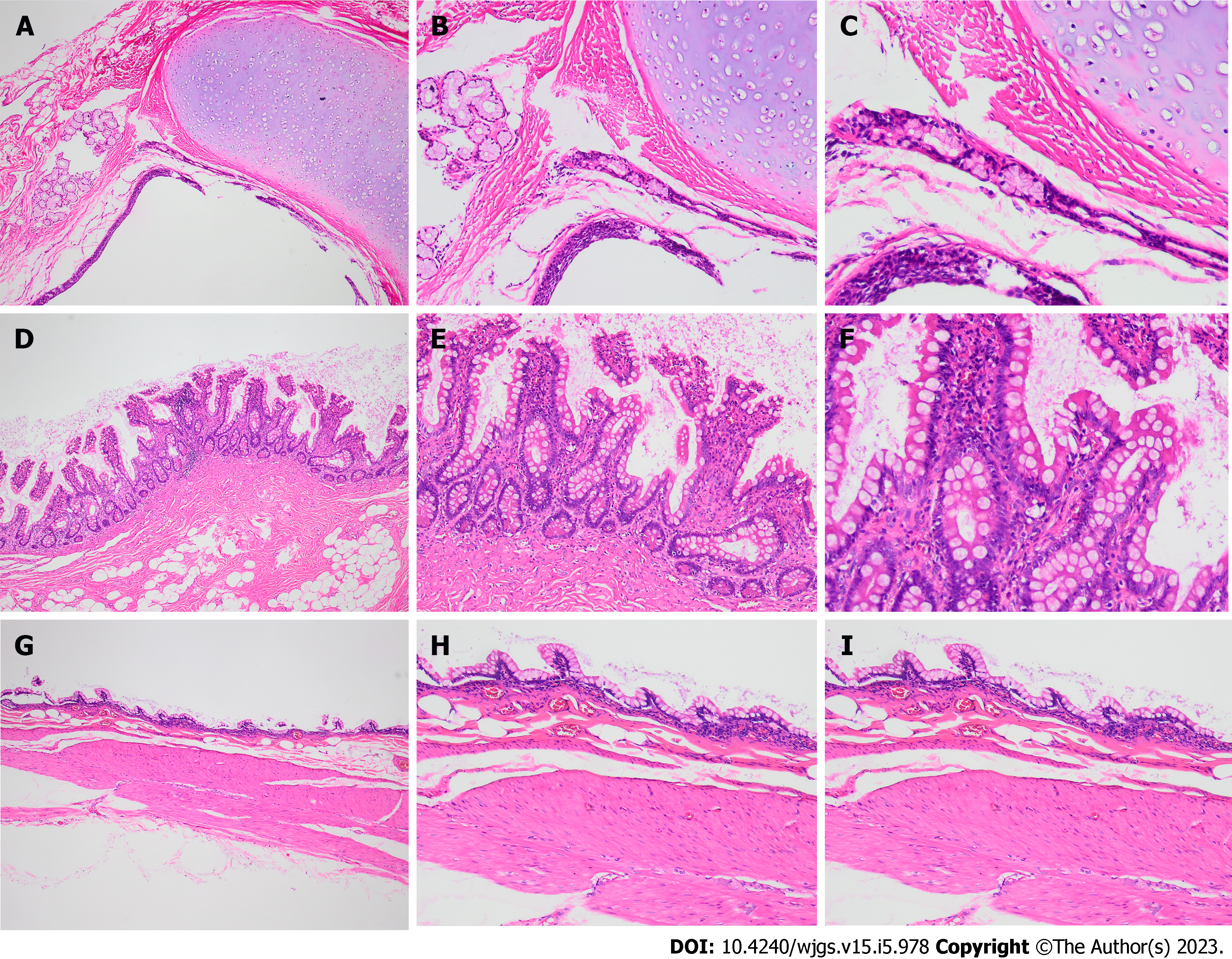
Figure 3 Postoperative pathological examination.
A-C: Hematoxylin-eosin stain of excised tissue showed mature cartilage tissue, small salivary glands and squamous epithelium; D-F: The intestinal mucosa and glands of the narrow intestinal cavity; G-I: The intestinal mucosa at the expansion site becomes thinner and the glands disappear. A, D and G: 40 × microscope; B, E and H: 100 × microscope; C, F and I: 200 × microscope.
- Citation: Xiong PF, Yang L, Mou ZQ, Jiang Y, Li J, Ye MX. Giant teratoma with isolated intestinal duplication in adult: A case report and review of literature. World J Gastrointest Surg 2023; 15(5): 978-983
- URL: https://www.wjgnet.com/1948-9366/full/v15/i5/978.htm
- DOI: https://dx.doi.org/10.4240/wjgs.v15.i5.978


