Copyright
©The Author(s) 2023.
World J Gastrointest Surg. May 27, 2023; 15(5): 812-824
Published online May 27, 2023. doi: 10.4240/wjgs.v15.i5.812
Published online May 27, 2023. doi: 10.4240/wjgs.v15.i5.812
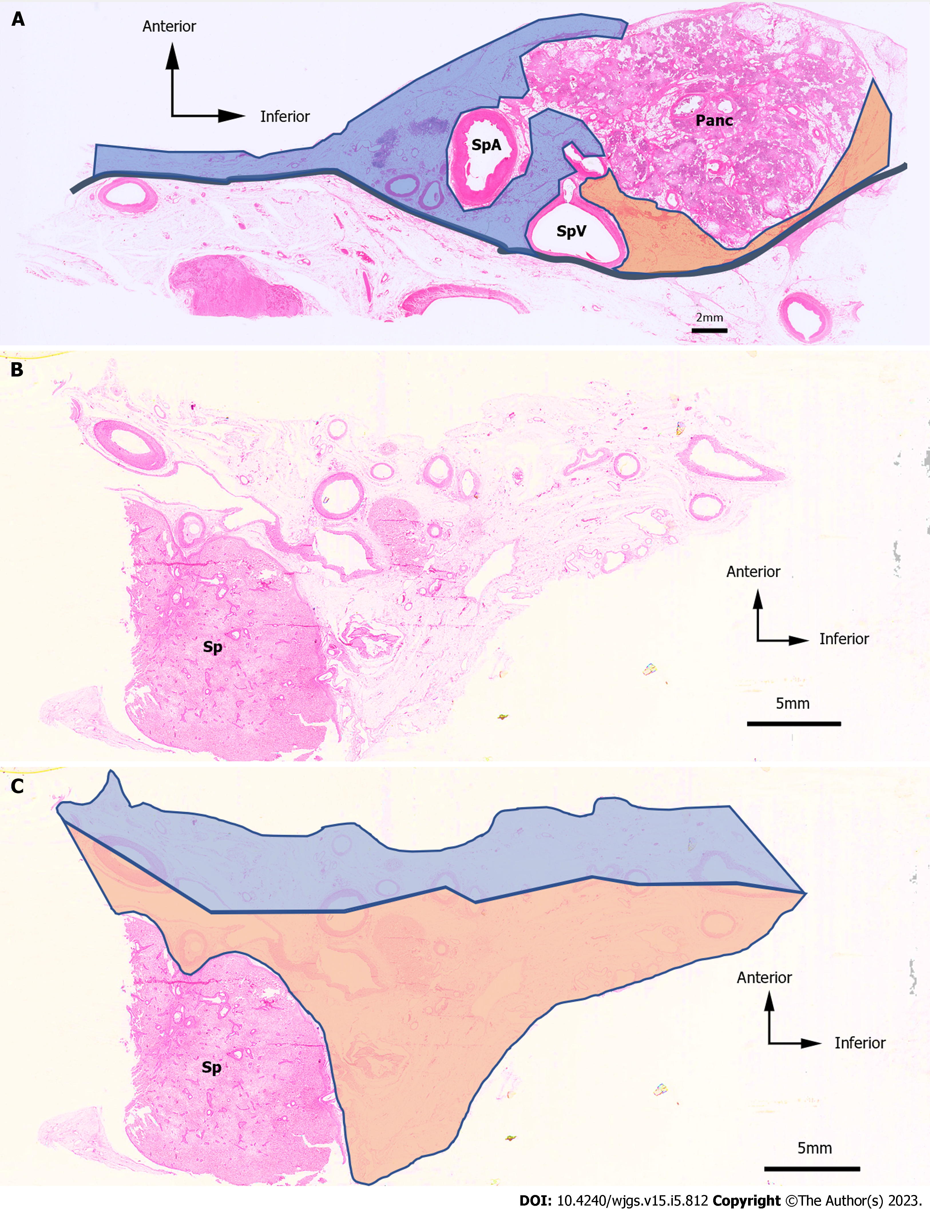
Figure 4 Hematoxylin & eosin-stained images and an example of the anterior side and the posterior side.
Hematoxylin & eosin (H&E)-stained images and an example of the anterior side and the posterior side. A: H&E-stained images of the No. 11 lymph node (LN) region in case 1. The anterior side is represented by the blue area, and the posterior side is represented by the orange area; B: H&E-stained images of the No. 10 LN region in case 4; C: The anterior side is represented as the blue area, and the posterior side is represented as the orange area. H&E: Hematoxylin and eosin; LN: Lymph node; Panc: Pancreas; SpA: Splenic artery; SpV: Splenic vein; Sp: Spleen.
- Citation: Umebayashi Y, Muro S, Tokunaga M, Saito T, Sato Y, Tanioka T, Kinugasa Y, Akita K. Distribution of splenic artery lymph nodes and splenic hilar lymph nodes. World J Gastrointest Surg 2023; 15(5): 812-824
- URL: https://www.wjgnet.com/1948-9366/full/v15/i5/812.htm
- DOI: https://dx.doi.org/10.4240/wjgs.v15.i5.812


