Copyright
©The Author(s) 2023.
World J Gastrointest Surg. May 27, 2023; 15(5): 812-824
Published online May 27, 2023. doi: 10.4240/wjgs.v15.i5.812
Published online May 27, 2023. doi: 10.4240/wjgs.v15.i5.812
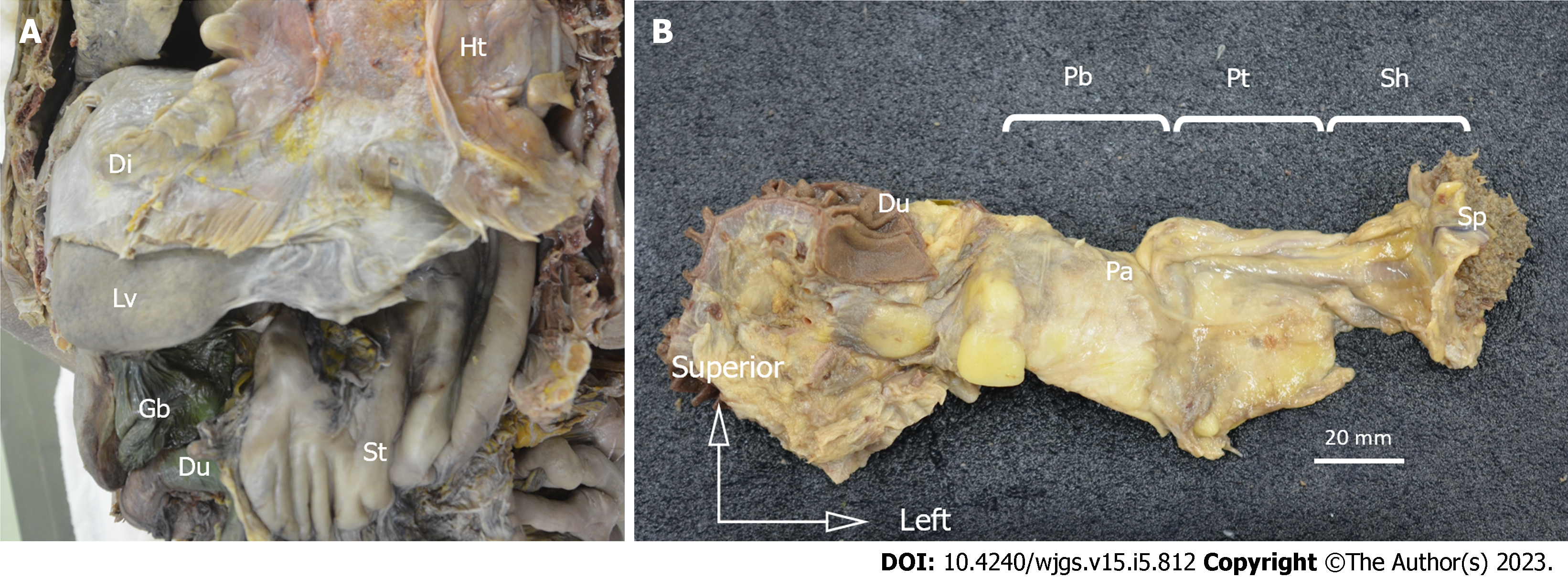
Figure 1 Resected specimens.
Resected specimens. A: Laparotomy was performed, and the esophagus, stomach, duodenum, spleen, pancreas, and vessels were resected in an en-bloc fashion; B: The duodenal bulb, horizontal leg, and lower bile duct were removed, and the duodenum, pancreas, spleen, and surrounding tissue were dissected from the retroperitoneum in an en-bloc fashion. Ht: Heart; Di: Diaphragm; Lv: Liver; St: Stomach; Du: Duodenum; Gb: Gallbladder; Pa: Pancreas; Pb: Pancreatic body; Pt: Pancreatic tail; Sh: Splenic hilum; Sp: Spleen.
- Citation: Umebayashi Y, Muro S, Tokunaga M, Saito T, Sato Y, Tanioka T, Kinugasa Y, Akita K. Distribution of splenic artery lymph nodes and splenic hilar lymph nodes. World J Gastrointest Surg 2023; 15(5): 812-824
- URL: https://www.wjgnet.com/1948-9366/full/v15/i5/812.htm
- DOI: https://dx.doi.org/10.4240/wjgs.v15.i5.812


