Copyright
©The Author(s) 2023.
World J Gastrointest Surg. May 27, 2023; 15(5): 799-811
Published online May 27, 2023. doi: 10.4240/wjgs.v15.i5.799
Published online May 27, 2023. doi: 10.4240/wjgs.v15.i5.799
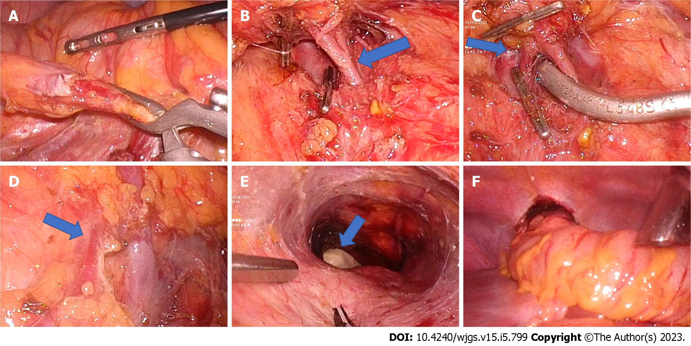
Figure 1 Laparoscopic mid-colon esophageal bypass.
A: Ileocolic pedicle dissected and bulldog clamp applied (marked with arrow); B: Middle colic artery dissected. Early division of the middle colic artery can be seen (marked with an arrow). Metal clips were applied on the right colic artery; C: Bulldog clamp applied on the middle colic artery proximal to the bifurcation. Middle colic vein draining to superior mesenteric vein seen (marked with arrow); D: The right colic vein (marked with arrow) joins with the gastrocolic trunk; E: Completion of retrosternal tunnel creation guided by assistant surgeon fingers; F: Transfer of colon conduit to the neck through the retrosternal tunnel.
- Citation: Kalayarasan R, Durgesh S. Changing trends in the minimally invasive surgery for corrosive esophagogastric stricture. World J Gastrointest Surg 2023; 15(5): 799-811
- URL: https://www.wjgnet.com/1948-9366/full/v15/i5/799.htm
- DOI: https://dx.doi.org/10.4240/wjgs.v15.i5.799


