Copyright
©The Author(s) 2023.
World J Gastrointest Surg. Mar 27, 2023; 15(3): 480-487
Published online Mar 27, 2023. doi: 10.4240/wjgs.v15.i3.480
Published online Mar 27, 2023. doi: 10.4240/wjgs.v15.i3.480
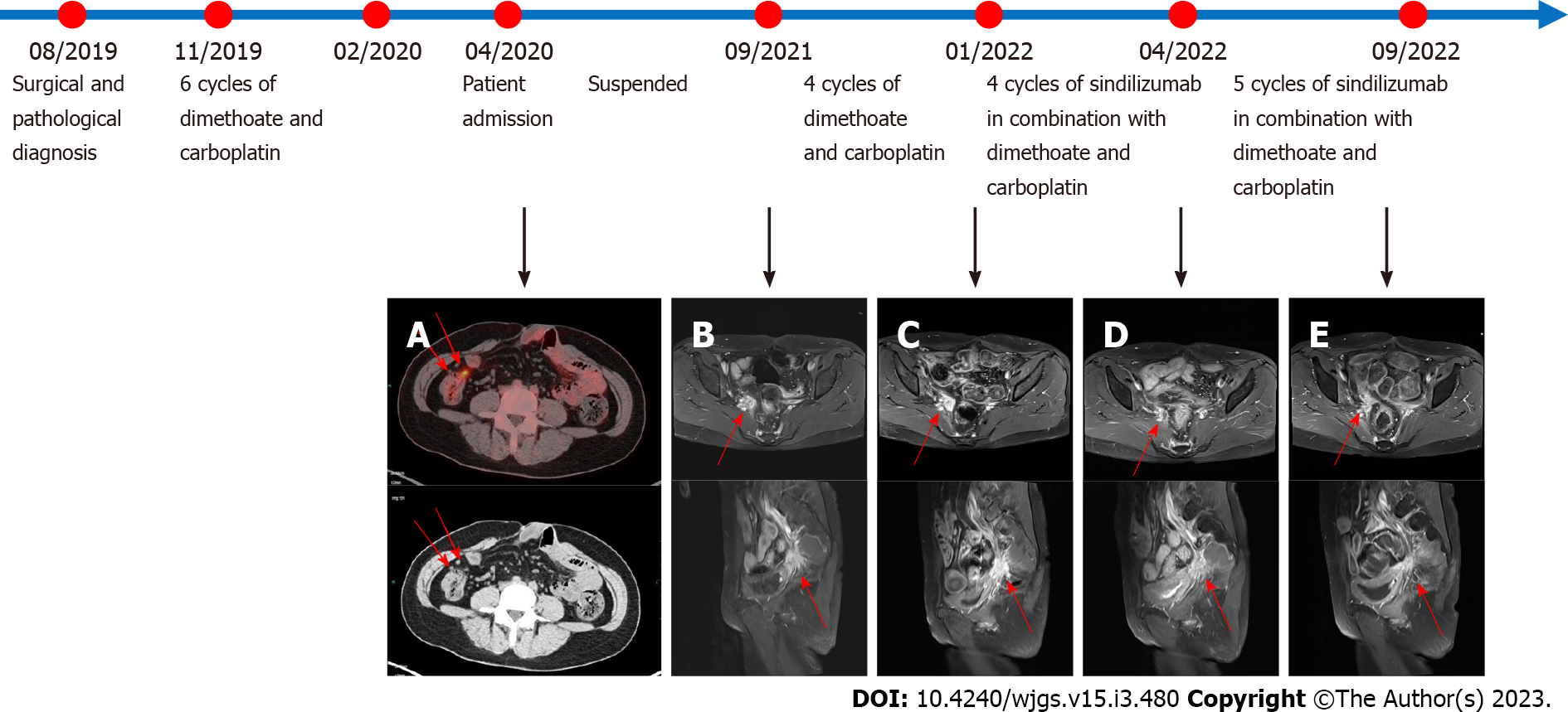
Figure 4 Pelvic magnetic resonance imaging scans before and after the use of sindilizumab and timeline of treatment.
Before the use of sindilizumab. A: The red arrows in the positron emission tomography-computed tomography images revealed multiple small nodular foci in the right pelvic wall and anterior sacral space with mildly increased fluorodeoxyglucose metabolism, which was considered to be due to tumor metastasis; B: The red arrow revealed an abnormal signal of about 22 mm × 24 mm mass is shown next to the right iliac vessels; C: After using 4 cycles of doxorubicin and carboplatin, the red arrow revealed the abnormal signal foci beside the right iliac vessels were slightly larger than before, with a size of 25 mm × 38 mm, which was considered to be caused by tumor recurrence. After the use of sindilizumab in combination with doxorubicin and cisplatin; D: The red arrow revealed the abnormal signal foci next to the right iliac vessels were significantly smaller than before, with a size of 17 mm × 12 mm; E: The red arrow revealed a small mass of abnormal signal foci, 17 mm × 12 mm in size, was observed next to the right iliac vessels, similar to the previous one. The timeline showed the whole process of diagnosis and treatment of this patient.
- Citation: Hu XC, Gan CX, Zheng HM, Wu XP, Pan WS. Immunotherapy in combination with chemotherapy for Peutz-Jeghers syndrome with advanced cervical cancer: A case report. World J Gastrointest Surg 2023; 15(3): 480-487
- URL: https://www.wjgnet.com/1948-9366/full/v15/i3/480.htm
- DOI: https://dx.doi.org/10.4240/wjgs.v15.i3.480


